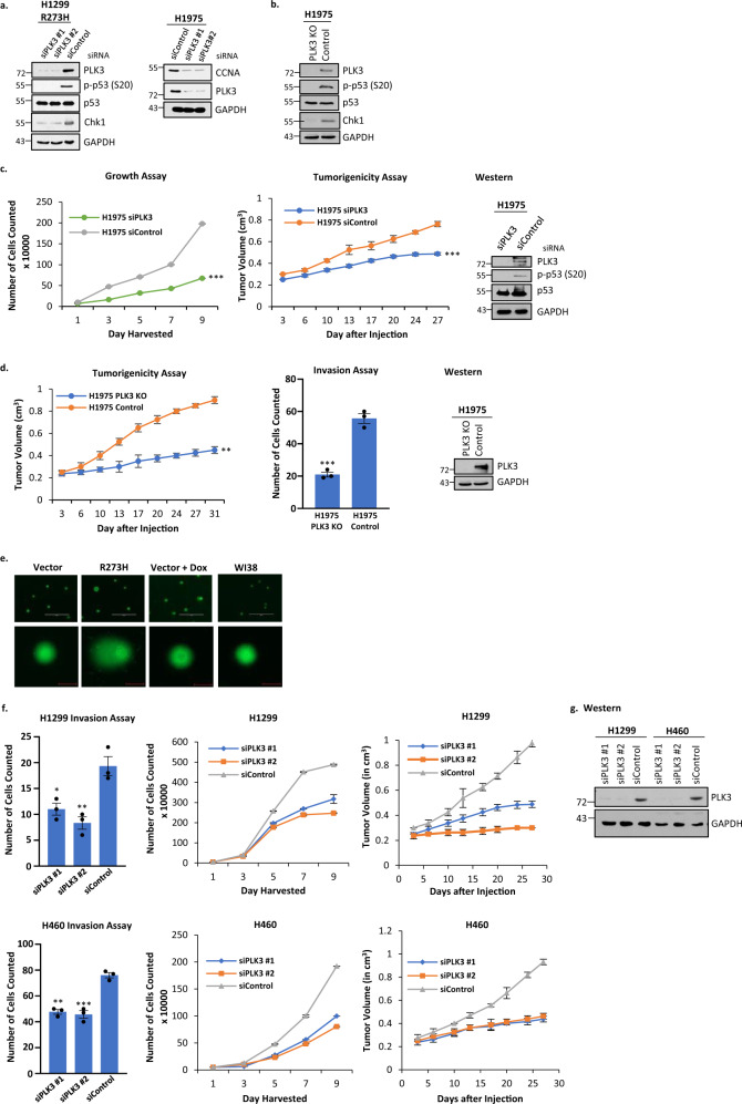
Fig. 7. PLK3 phosphorylates S20 of mutant p53 and affects cell growth, tumorigenicity, and invasion in cells harboring GOF mutant p53.
a H1299 cells expressing p53-R273H were transfected with siRNA specific for PLK3 (or scrambled control) and after incubation cells were harvested to analyze PLK3 levels and phosphorylation at S20 of p53. Data presented show that treatment of H1299 cells expressing p53-R273H with siPLK3 decreases the PLK3 protein concordant with loss of phosphorylation at S20; concomitantly the level of Chk1, a mutant p53 inducible gene, was reduced. An additional mutant p53 inducible gene, CCNA, was also reduced after PLK3 reduction in H1975. These observations suggest that S20 phosphorylation modulates transactivation of mutant p53. b A PLK3 knockout (PLK3 KO) cell line was generated to further prove that PLK3 modulates mutant p53’s transactivation via phosphorylation of S20. PLK3 was knocked out in the H1975 lung cancer cell line using CRISPR (region of deletion located to Chr1 44803749-44805264). Cell extracts from H1975 PLK3 KO and corresponding CRISPR control (Control) were harvested to analyze PLK3, p-p53 (S20), and Chk1 levels as shown. Data recapitulated those seen in the siRNA experiment shown in a. c H1975 lung cancer cells were transfected with siRNA against PLK3 (or scrambled control) and growth and tumorigenicity assays were performed. Data are presented as the mean ± SEM (n = 3 for growth assay, n = 4 for tumorigenicity assay). A two-sided Student’s t-test was performed; Growth assay ***p = 1.8E–7, Tumorigenicity assay ***p = 5.2E–5. In both assays, siPLK3 reduced the oncogenic potential of the cell line. d A PLK3 knockout (KO) cell line was generated to further prove that PLK3 can be targeted to reduce the oncogenicity of lung cancer cells harboring mutant p53. PLK3 was knocked out in the H1975 lung cancer cell line using CRISPR (region of deletion located to Chr1 44803749-44805264) and tumorigenicity and invasion assays were performed. Data are presented as the mean ± SEM (n = 4 for tumorigenicity assay, n = 3 for growth assay). A two-sided Student’s t-test was performed; Invasion assay ***p = 0.0004, Tumorigenicity assay **p = 0.007. Both assays showed that the tumorigenic and invasive properties are severely reduced when PLK3 is knocked out. Western blots show levels of PLK3/p53/phospho-p53. e A comet assay was performed on H1299 cells stably transfected with empty vector and expressing p53-R273H. H1299 cells stably transfected with empty vector and treated with doxorubicin were used as a positive control while untreated WI38 cells were a negative control. (Scale bar: 400 µM, top row; 40 µM bottom row). f Invasion, growth, and tumorigenicity assays were performed on H1299 and H460 cells after PLK3 was knocked down using siRNA. Data are presented as the mean ± SEM (n = 3 for invasion and growth assays, n = 4 for tumorigenicity assay). g Western blot analysis of cells used in f showing efficient knockdown of PLK3 after siRNA treatment. *p-value < 0.05, **p-value < 0.01, and ***p-value < 0.001. NS no statistically significant difference from control.

