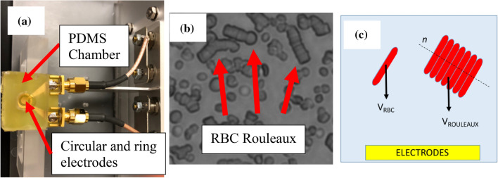
Figure 1.
Blood samples were housed in a PDMS chamber and their electrical impedance was measured using circular and ring electrodes as shown in (a). As blood is left at hemostasis in autologous plasma, RBCs aggregate to form ‘rouleaux’. A representative microscopic picture of RBC rouleaux is shown in (b). As the sedimentation velocity of a particle in low Reynolds number regime is dependent on its radius squared, RBC Rouleaux sediment faster than RBCs. A schematic depicting the cross-section of the chamber is shown in (c).

