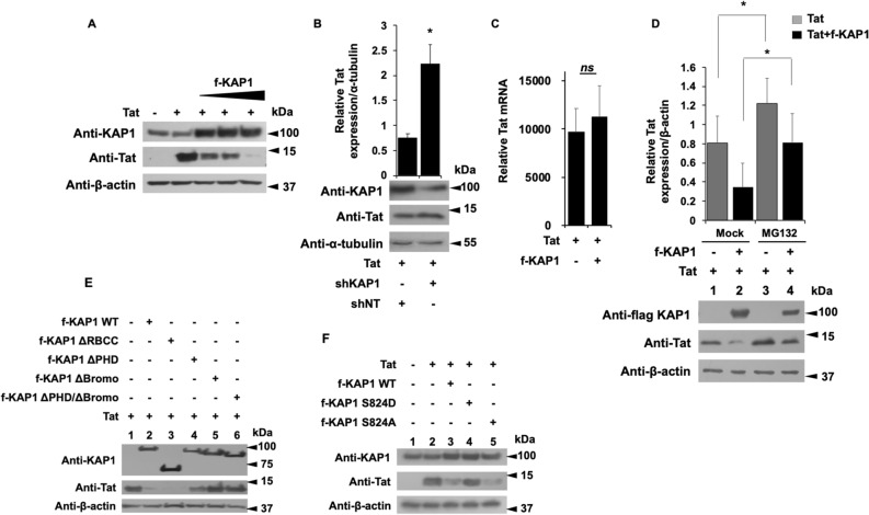
Figure 7.
The Bromo domain of KAP1 promotes Tat degradation via the proteasome pathway (A,B,E,F) HEK cells were transfected with the indicated vectors. 48 h later, the nuclear proteins were analyzed by Western blot for the presence of the indicated proteins. (B) Tat expression level whose relative quantification to the α-tubulin was carried out using the image J software. (C) The RNA extracts from HEK cells transfected with the indicated plasmids were submitted to RT-qPCR experiments against Tat. The relative mRNA level of Tat was normalized to the GAPDH gene. (D) HEK cells were transfected with the indicated vectors. After 18 h of transfection, the cells were treated or not with 50 μM of MG132 for 6 h. 24 h post-transfection, the total protein extracts were analyzed by Western blot for the presence of KAP1 and Tat. Tat expression level was quantified relatively to β-actin expression, using image J software. A t-test was performed on 3 (B,C) and 5 (D) independent experiments P (*P < 0.05, **P < 0.01, ***P < 0.001). Full-length blots are presented in Supplementary Figure 7.

