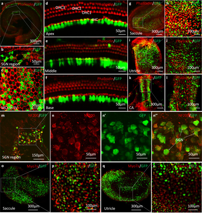
Fig. 1. Confocal immunofluorescent images of whole-mount preparations of cochleae and vestibules in WT mice at 1 month after AAV1-CB7-GFP delivery via PSCC injections at P0–P2 (green: GFP; n = 6 in each group).
a A representative low-magnification image of an apical middle turn. b A high-magnification image of the spiral ganglion neuron (SGN) tissue. c A high-magnification image of marginal cells (MCs) in the stria vascularis (SV). d–f High-magnification images of the apex, middle, and base of the basilar membrane. g–j Representative low- and high-magnification images of saccule and utricle. k, l Representative low- and high-magnification images of the crista ampullaris (CA). m Representative low-magnification images of SGNs co-labeled with antibodies against NF200 (red) and GFP. n, n” Representative high-magnification images of SGNs (boxed area in m) labeled with antibodies against NF200 (n, red) and GFP (n’), n” is the superimposed images of n and n’. o–r Representative low- (o, q) and high (p, r) magnification images of saccule and utricle co-labeled with antibodies against Myo7a (red) and GFP.

