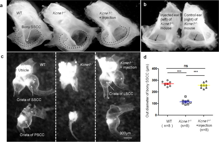
Fig. 3. Morphology of semicircular canals and vestibular membranous labyrinths in WT, untreated Kcne1−/− mice, and treated Kcne1−/− mice at P30 (n = 8 in each group).
a Comparison of the gross morphologies of bony semicircle canals in WT, untreated Kcne1−/− mice, and treated Kcne1−/− mice. Border of bony superior semicircular canal (SSCC) is outlined by dashed lines. b Gross morphologies of bony posterior semicircular canal (PSCC) in left and right ear of treated and untreated ears of the same Kcne1−/− mouse. Arrows point the region where the diameters of the semicircular canals were measured. c Gross morphologies of vestibular membranous labyrinths in WT, untreated Kcne1−/− ear, and treated ears of Kcne1−/− mice. d Quantification of the outer diameter of bony SSCC in WT, untreated Kcne1−/− mice, and treated Kcne1−/− ears as labeled (n = 8 in each group, p < 0.0001 in WT ears, and p < 0.0001 in treated Kcne1−/− ears, comparing to untreated Kcne1−/− ears, two-sided Student’s t tests). ***: p < 0.001. Data are shown as mean ± SEM. Source data are provided as a Source data file.

