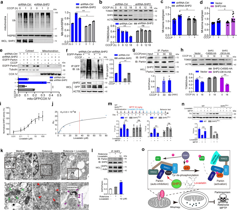
Fig. 1.
SHP2-mediated mitophagy enhanced by lovastatin in neuronal cells alleviates parkinsonism in mice. a Immunoblot analysis of the ubiquitination of mitochondria in shRNA-SHP2 or shRNA-Ctrl SH-SY5Y cells treated with 10 μM CCCP for indicated times. b Immunoblot analysis of TOM20 degradation in shRNA-SHP2 or shRNA-Ctrl SH-SY5Y cells in the presence of 10 μM CCCP for indicated times. c, d Quantification of mt-Keima (red/green) in SH-SY5Y cells with SHP2 knockdown or overexpression. The cells were treated with 10 μM CCCP for 1 h and then imaged with 458 nm (green) or 543 nm (red) light excitation. e Immunoblot analysis of Parkin’s mitochondrial translocation in shRNA-SHP2 or shRNA-Ctrl SH-SY5Y cells transfected with EGFP-Parkin for 24 h in the presence of 10 μM CCCP for 1 h. f Immunoblot analysis of Parkin ubiquitination in HeLa cells co-transfected with shRNA-SHP2 and EGFP-Parkin for 24 h followed by 10 μM CCCP treatment for 1 h. g Co-immunoprecipitation (Co-IP) analysis for the interaction of SHP2 with Parkin in SH-SY5Y cells treated with 10 μM CCCP for indicated times. h Immunoblot analysis of TOM20 expression from SH-SY5Y cells transfected with vector or SHP2-C459S or SHP2-D61A as indicated in the presence of 10 μM CCCP for 18 h. i The purified SHP2 protein was incubated with different doses of lovastatin and then the SHP2 enzyme activity was examined. j Interaction between lovastatin and SHP2 was determined via SPR analysis. k SH-SY5Y cells were pretreated with 10 μM lovastatin for 3 h followed by the addition of 30 μM rotenone for 6 h. Cells were collected for transmission electron microscopy assay. Green arrow, normal mitochondria; red arrow, swollen mitochondria; purple arrow, the damaged mitochondria localized near a lysosome in the autophagolysosome. l Co-IP analysis of Parkin and SHP2 in SH-SY5Y cells pretreated with 10 μM lovastatin for 3 h followed by the addition of 30 μM rotenone for 6 h. m Overview of the experimental design and behavioral tests for WT and SHP2TH−/− mice treated with MPTP and lovastatin. n The expressions of TH in the striatum of each group were measured using Western blot analysis. o The graphic illustration of the mechanism of lovastatin-driven SHP2-mediated dephosphorylation of Parkin in promoting mitophagy in neuronal cells and alleviating parkinsonism in mice. Upon the initiation of mitochondrial damage in SH-SY5Y neuronal cells, SHP2 translocates to mitochondria, where it directly interacts with Parkin and promotes its E3 ligase activity via decreasing Parkin phosphorylation, increasing mitophagy as well as neuronal cell survival. Lovastatin promotes SHP2/Parkin-mediated mitophagy and exerts neuroprotective effects in MPTP-challenged mice. Data are representative of three independent experiments (mean ± SEM). *P < 0.05, **P < 0.01 vs. indicated

