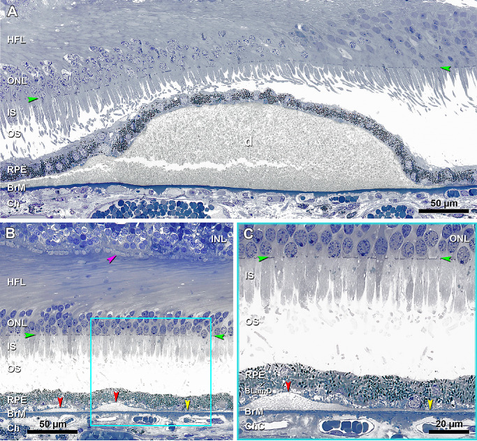
Figure 3.
BLinD and soft drusen are two forms of the same deposit, and pre-BLinD is the precursor. (A) A large soft druse (d) is located in the sub-RPE-basal laminar space, with a granular internal structure that stains gray and continuous with BLinD to the left. The crack is artifact. Seventy-six-year-old woman. (B) Undulating layer of BLinD (red arrowheads) and flat layer of pre-BLinD (yellow arrowhead) are observed in the same sub-RPE-basal laminar space and have a similar internal structure and staining characteristics as soft drusen. Cone pedicles with dark staining synaptic complexes (fuchsia arrowhead) indicate good tissue fixation. Teal frame shows BLinD and pre-BLinD magnified in panel C. Artifactual retinal detachment was digitally approximated to the RPE (Photoshop). (C) BLinD and pre-BLinD consist of finely granular gray-staining material (red and yellow arrowheads, respectively). Pre-BLinD stains darker than BLinD. Ninety-year-old woman. Ch, choroid; HFL, Henle fiber layer; IS, inner segment; ONL, outer nuclear layer; OS, outer segment. Green arrowheads, external limiting membrane.

