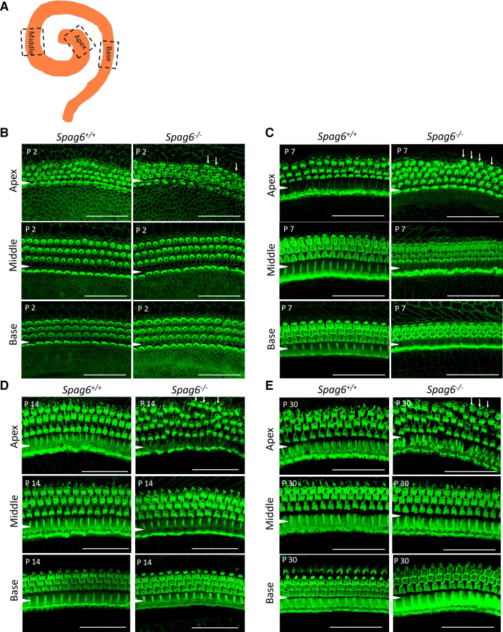
Figure 2.
Spag6 inactivation alters cellular arrangement. Whole-mount preparations were prepared at postnatal day 2 (P2), P7, P14, and P30. A: diagram of observed apical, middle, and basal turn. B: at P2, in the Spag6+/+ mice, irregularly arranged hair cells could be seen occasionally because of immaturity. There were more than three rows of outer hair cells (OHCs) in the apical turn of Spag6−/− mice (white arrows). C−E: at P7 and after, the Spag6+/+ mouse inner hair cells (IHCs) displayed a normal arrangement in apical, middle, and basal turns. However, in the P7, P14, and P30 Spag6−/− mice, there still was a disrupted arrangement of OHCs, mainly in the apical turn indicating defects in development. (white arrows in C–E). A white arrowhead is located in the region between the IHCs and OHCs. Scale bars: 30 µm.

