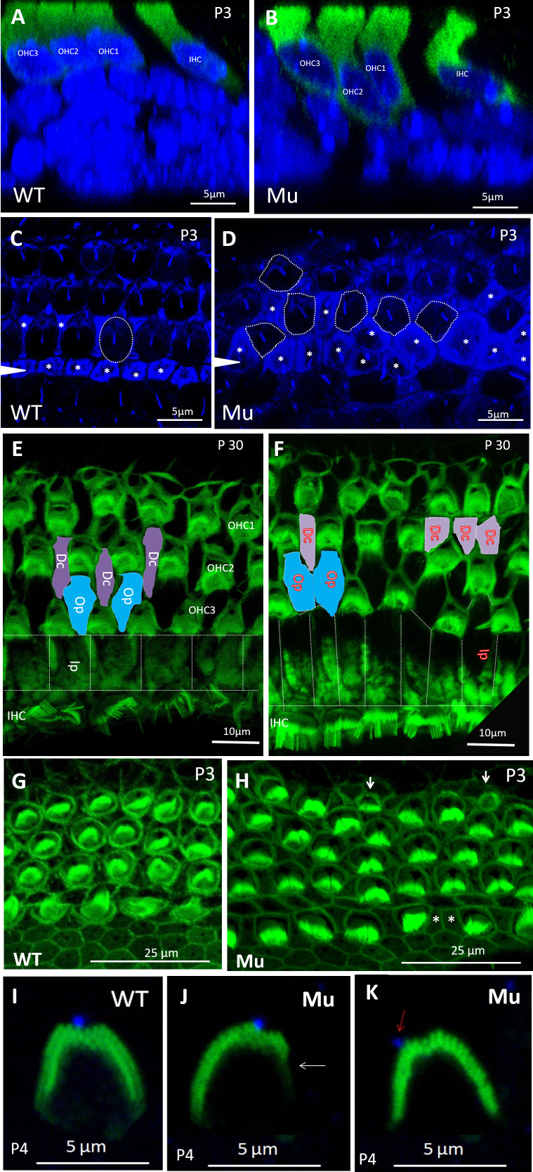
Figure 3.
Spag6 inactivation alters cell shapes and cellular arrangement. Optical sectioning and whole-mount preparations were observed. Mice of any sex were used. A and B: nuclei (blue) of Spag6+/+ OHCs (green) at P3 were uniformly positioned (A); however, Spag6−/− OHCs were positioned at variable heights in the apical-basal axis of the sensory epithelium (B). HCs were stained by antimyosin 7a antibody. C and D: acetylated-α-tubulin (blue) marked microtubule-rich structures like supporting cells, kinocilia, and the cuticular plate of HCs. Cochlear specimens from Spag6+/+ mice at P3 displayed precisely aligned hair cell rows. Hair cells showed oval shape (C; dotted circle) separated by regularly shaped supporting cells (asterisks in C). In contrast, cochlea of the Spag6−/− mice showed a disordered arrangement and irregular shape of OHCs (dotted circles in D) and supporting cells (asterisks in D). E and F: cochlear apex (phalloidin, green) of Spag6+/+ mice at P30 showed a normal arrangement of OHCs and supporting cells, as illustrated (blue and purple block in E). However, in the Spag6−/− mouse, some supporting cells were adjacent without intervening HCs (blue and purple block in F). G and H: compared with the Spag6+/+ mice, cochlea of Spag6−/− mice showed abnormally shaped OHCs and supporting cells in the apex. Some smaller OHCs with or without stereocilia bundles could be found in the Spag6−/− mice (arrows in H). I, J, K: in the basal turn of Spag6+/+ cochlea at P4, the stereocilia bundle was “V”-shaped and basically symmetrical, the kinocilium (blue) was located at the apex of the “V” (I). In contrast, some Spag6−/− hair cell stereocilia bundles had a defective “V” shape and were asymmetrical (arrow in J), the kinocilium was not at the top of “V” (arrow in K). Dc, Deiter’s cell; IHC, inner hair cells; Ip, inner pillar cell; Mu, Spag6−/−; OHC, outer hair cells; Op, outer pillar cell; WT, Spag6+/+; WT, wild type.

