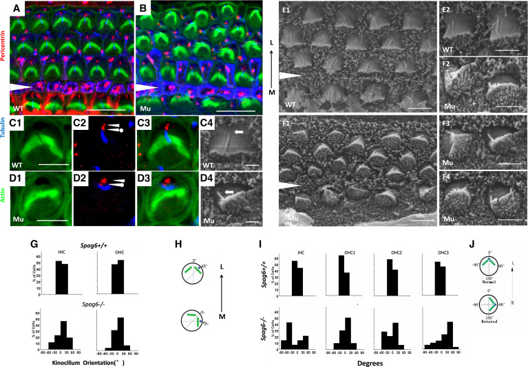
Figure 4.
The position of kinocilia and basal bodies are affected in Spag6−/− mice. A–F: confocal projections of cochlear whole mounts and scanning electron micrographs of hair cells from control and Spag6−/− animals at postnatal day 1 of either sex. A and B, C1–C3, and D1–D3: cochlear whole mounts were stained with a pericentrin antibody (red) and an acetylated-α-tubulin antibody (blue) to mark centrioles within the basal bodies and kinocilium, respectively. The cytoskeleton was labeled with phalloidin (green). In a Spag6+/+ mouse, the maternal centriole (C2, arrowhead with a dot) was located at the base of the kinocilium (C2, blue), and the daughter centriole (C2, arrowhead) was positioned medially to the maternal centriole. This pair of centrioles was aligned along the mediolateral axis, and the kinocilium was at the vertex of the “V”-shaped stereocilia bundle (C4, arrow). In the Spag6−/− mice, the maternal centriole (D2, arrowhead with a dot) and daughter centriole (D2, arrowhead) were no longer aligned mediolaterally. The kinocilium (D2, blue) was apart from the vertex of the stereocilia bundle, disoriented, and no longer aligned along the mediolateral axis (D4, arrow). E and F: scanning electron microscopy (SEM) examination of wild-type (E1 and E2) and Spag6−/− mice (F1–F4) hair cells of apical turn at postnatal day 1. Spag6+/+ hair cells showed normal polarity (E1), and the kinocilium was in the vertex of the V-shaped stereocilia bundles (E2, arrows). In the Spag6−/− mice, the polarity of hair cells was disturbed (F1). The intrinsic polarity of some hair cells was affected, showing malformations (F2–F4), and kinocilia were positioned at the periphery in the same general direction as the disoriented bundles (F2–F4, arrows). The black arrowheads (in A, B, E1, F1) mark the support cell region between the inner hair cells (IHCs) and outer hair cells (OHCs). G−J: examination of the relationship between kinocilium position and bundle rotation in hair cells of the apical turn according to SEM observations at P1. In Spag6+/+ mice (G), there was a correlation between the kinocilia position and the apex of stereocilia bundles. In Spag6−/− mice (G), hair cell bundle and kinocilia were frequently mislocalized and distorted. The degrees between kinocilia position and the apex of stereocilia bundles varied from −60°to +60° (Spag6+/+, n = 160; Spag6−/−, n = 142). Diagrams (H) illustrate measurements of the relationship between kinocilium position and bundle rotation (degree). I: In Spag6−/− mice, the degrees of stereocilia bundle orientation were more than ±30°, compared with the Spag6+/+ mice. Diagrams (J) illustrate measurement of the bundle rotation (degrees). White triangle is located in the region between the outer and inner hair cells. L, lateral; M, medial. Scale bars: A and B, 10 μm; C−F, 5 μm.

