Figure 6.
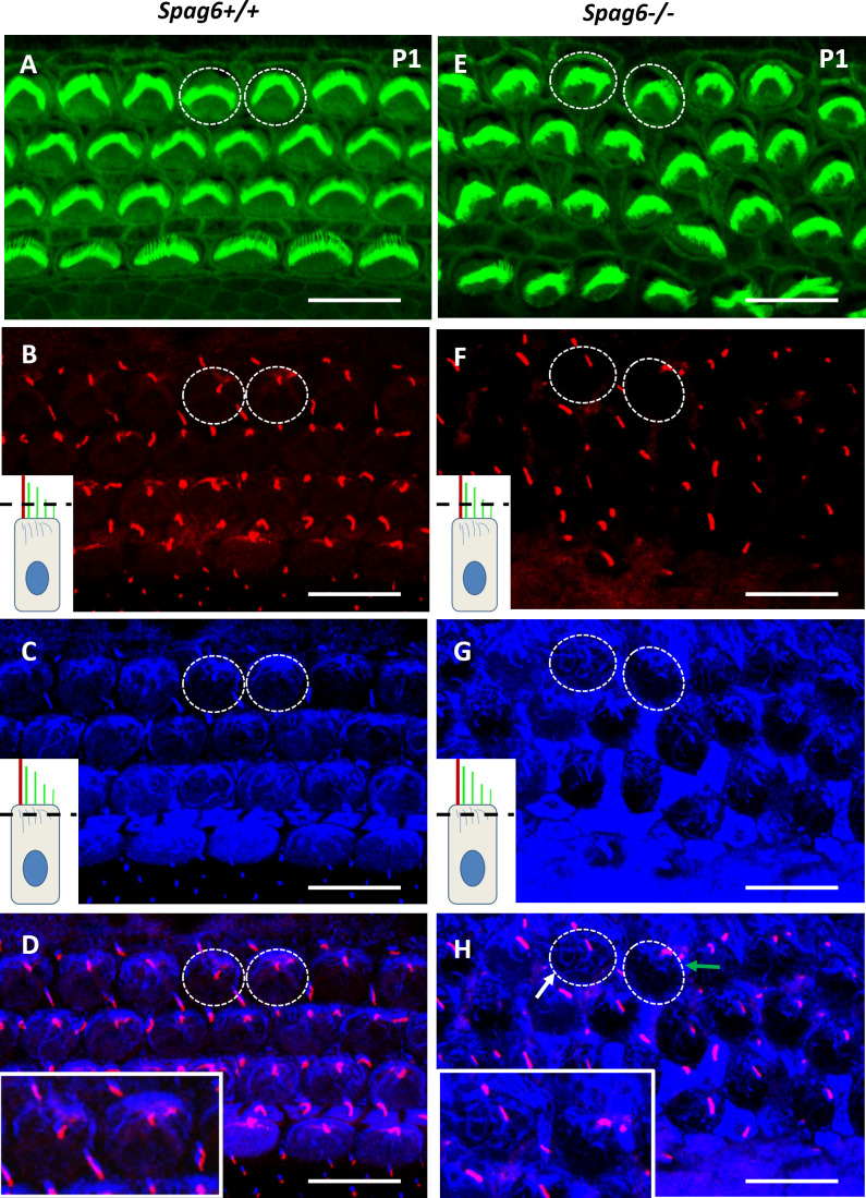
The apical cytoskeleton is altered in Spag6−/− mice. Whole-mount preparation were immunostained for actin (green) and acetylated-α-tubulin (white) to visualize stereocilia, kinocilia, and cytoplasmic microtubules. Confocal images were taken from the kinocilia level (see inset diagram, B and F) to the apical microtubule level (C and G). B and C, F, and G were overlapped respectively, to see the apical microtubule system and the kinocilium in each hair cell at the same time (D and H). The inset images in B, C, F, and G showed the scan level (black dotted line). Two hair cells were selected randomly in A and E appearing as a dotted-line circle and magnified for better visualization (D and H, see inset.). D: in the Spag6+/+ mice, the apical microtubules (blue) radiated along the periphery of the cell cortex from the base of kinocilium (red) where basal bodies were located. H: in the Spag6−/− mice, the apical microtubules were disturbed and distributed disorderly in the cuticular plate (white arrpw, left dotted circle). The kinocilium was dislocated (red), which is consistent with the misorientation of the microtubule network (green arrow, right dotted circle). Mice of either sex were used. Scale bar: A and B; 10 μm.

