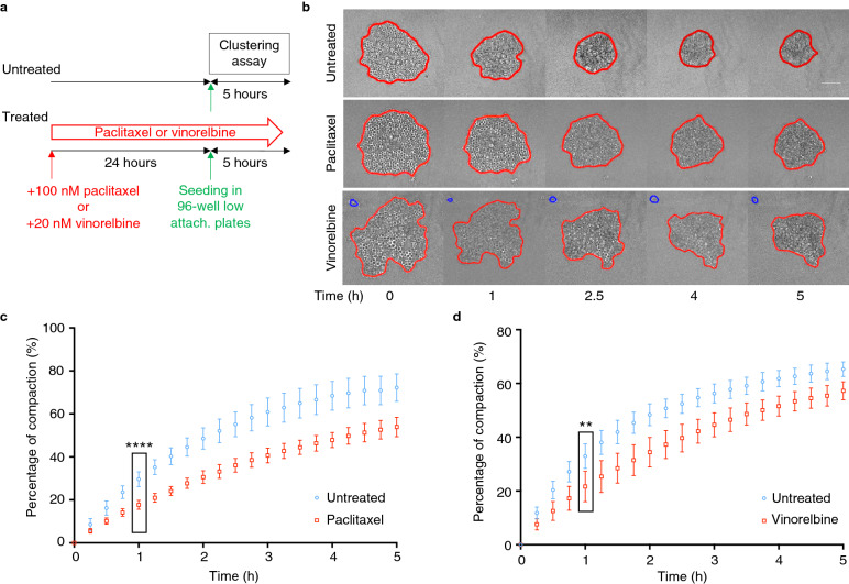
Fig. 1.
Aggregation is compromised in MCF-7 cells incubated with paclitaxel or vinorelbine. a Schematic representation of the experimental set-up. MCF-7 cells incubated or not (untreated) with paclitaxel (100 nM) or vinorelbine (20 nM) for 24 h were seeded in 96-well low-attachment (low-attach.) plates and monitored by video-microscopy for 5 h (clustering assay) (adapted from [15]). b Representative transmitted light microscopy images of cell aggregation at the indicated time points. Segmentation (red line) was performed using a dedicated MATLAB software. Blue lines correspond to isolated cells. c, d An automated image processing procedure was used to measure the aggregate area during the assay in the presence of paclitaxel (c) or vinorelbine (d) and the percentage of compaction was calculated from the normalized area variation relative to the initial time point. Data correspond to the mean ± SD of 48 aggregates/condition from 3 independent experiments. **P < 0.01; ****P < 0.0001 (Mann–Whitney non-parametric test)

