Figure 9.
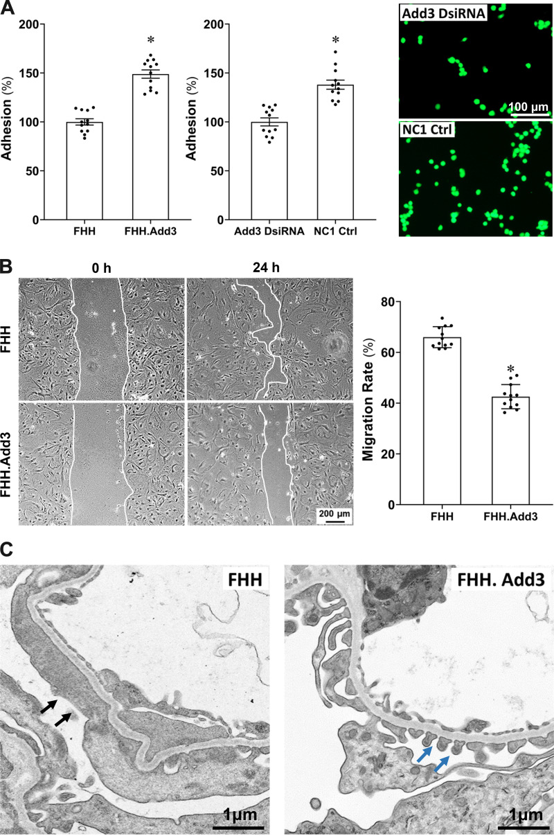
Reduced γ-adducin (ADD3) expression is associated with diminished podocyte adhesion, enhanced podocyte migration, and podocyte foot process effacement (FPE) in fawn-hooded hypertensive (FHH) rats. A: podocyte adhesion was decreased in male FHH compared to FHH.Add3 rats (left) and in mouse podocyte transfected with Add3 Dicer-substrate short interfering RNA (DsiRNA) vs. NC1 control (Ctrl; middle). Representative images of fluorescent adhesive cells are presented on the right. B: representative images (left) and quantitation (right) demonstrate that migration rate of primary podocytes was significantly higher in male FHH than FHH.Add3 rats. C: representative images show that knockin of wild-type Add3 improved podocyte ultrastructure and rescued FPE observed in age-matched FHH rats. Black arrows denote the area of FPE in FHH rats. Blue arrows denote intact slit diaphragms in FHH.Add3 rats. Magnification: ×32,100. Experiments using conditionally immortalized mouse podocytes and primary podocytes were repeated 3–4 times in triplicate in each experiment. Podocytes were isolated from 4–6 male rats in each strain. Mean values ± SE are presented. *P < 0.05 from the corresponding values in age-matched FHH rats or NC1 negative controls.

