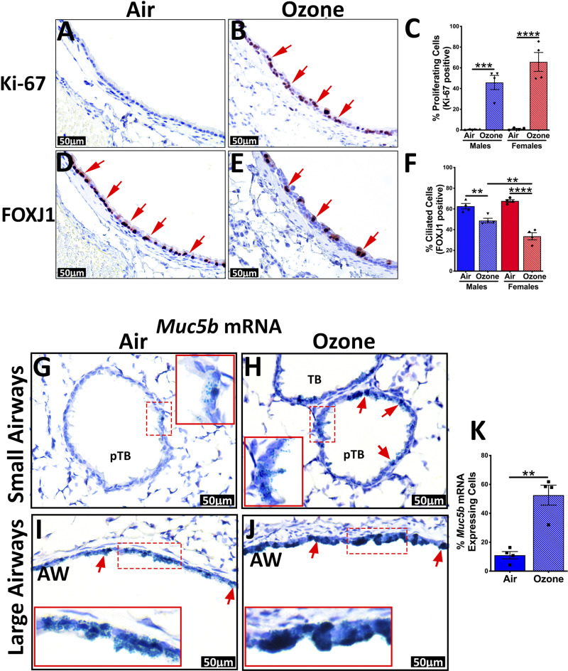
Figure 4.
Immunohistochemical and in situ RNAscope staining of airways for epithelial remodeling-associated changes. Representative photomicrographs of Ki-67 stained lung sections from air-exposed (A) and ozone-exposed mice (B). C: percentage of Ki-67 stained cells in the first-generation airways from air- and ozone-exposed mice. Representative photomicrographs of FOXJ1-stained lung sections from air-exposed (D) and ozone-exposed mice (E). F: percentage of FOXJ1-stained cells in the first-generation airways from air- and ozone-exposed mice. Error bars represent means ± SE **P < 0.01, ***P < 0.001, ****P < 0.0001 using one-way ANOVA followed by the Tukey multiple-comparison post hoc test. (n = 4 per group). G–J: mRNA expression of Muc5b in airways of air-exposed and ozone-exposed mice was detected by RNAscope assay. Representative photomicrographs depicting Muc5b mRNA signal (green dots, as well as green-stained cells, red arrows) air-exposed (G; small airways, I; large airways) and ozone-exposed mice (H; small airways, J; large airways). Inset outlined with red solid box is the higher magnification of what is depicted in the red dashed box. K: percentage of Muc5b mRNA-expressing cells in the smaller airways from air- and ozone-exposed mice. AW, 1st generation airway; pTB, preterminal bronchiole; TB, terminal bronchiole.

