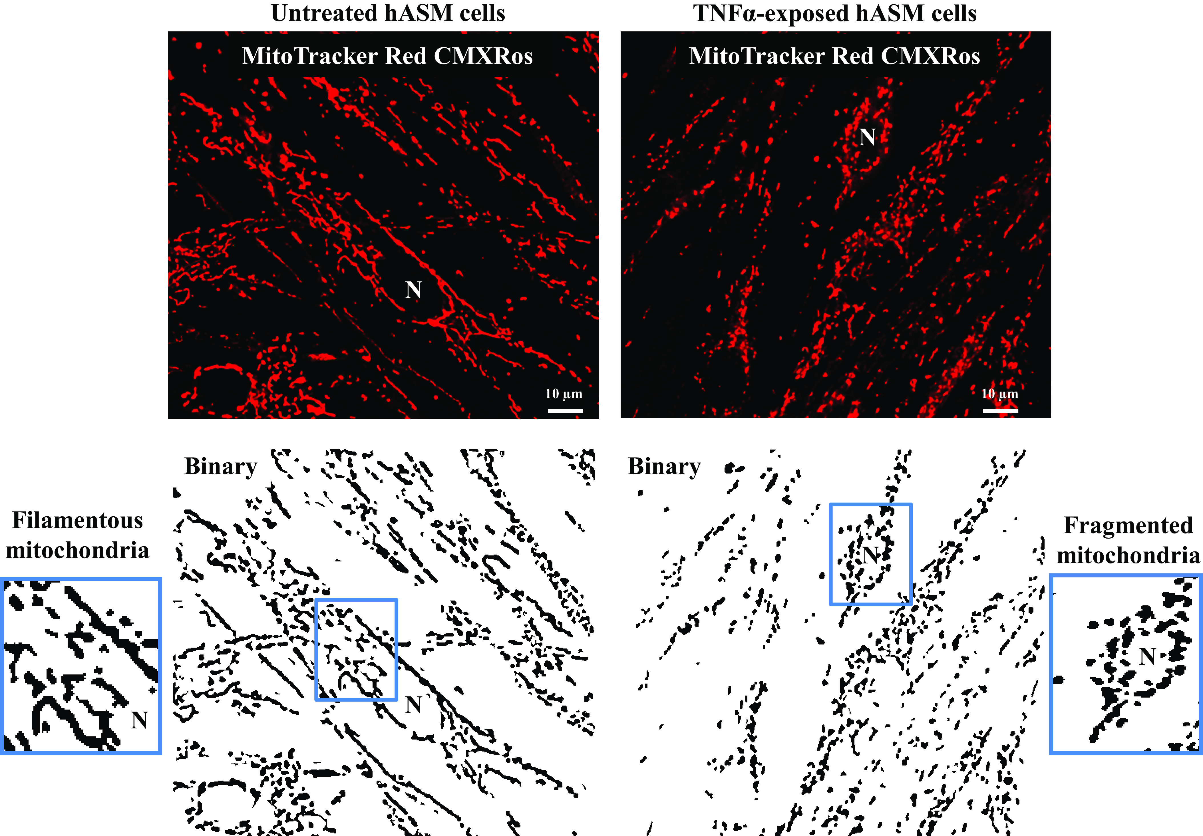
Figure 2.

Representative 2-dimensional (2-D) fluorescent confocal images of human airway smooth muscle (hASM) cells untreated or treated with TNFα loaded with MitoTracker Red CMXRos to visualize mitochondria and their corresponding binary images showing mitochondrial morphology. In untreated hASM cells, mitochondria appear filamentous, while in TNFα-treated hASM cells, mitochondria appear fragmented or globular. N, nucleus.
