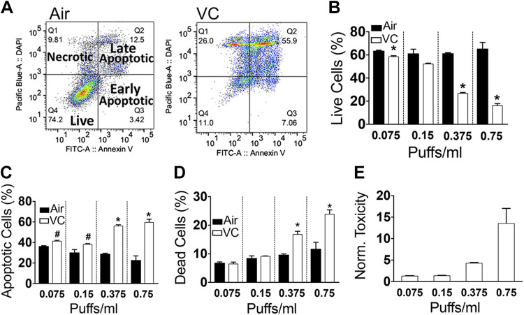
Figure 2.
Quantification of annexin V staining in HL-1 cells with flow cytometry. A: flow cytometry analysis of live, apoptotic, and necrotic HL-1 cells cultured for 48 h with 0.75 puffs/mL air or 0.75 puffs/mL vanilla custard (VC) e-vapor extract. B–D: flow cytometry quantification of the percentage of live (B), apoptotic (C), and necrotic (D) cells, in air or vanilla custard e-vapor bubbled medium at 0.075, 0.15, 0.375, and 0.75 puffs/mL. E: toxicity index normalized to that of air. n = 3 in each condition. #P < 0.05 vs. air; *P < 0.01 vs. air, t test.

