Figure 4.
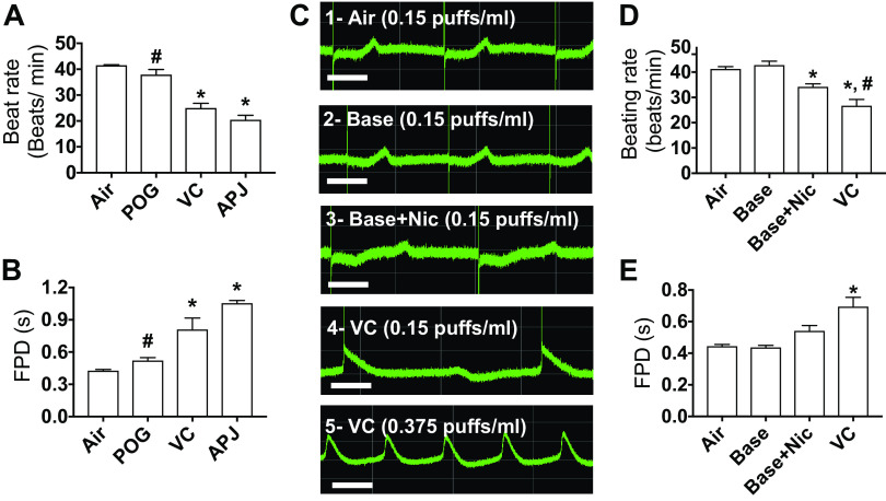
Effects of 24 h, 0.15 puffs/mL e-vapor exposure on beating rate, and corrected field potential duration in human iPSC-derived cardiomyocytes. Quantification of beating rate (A) and field potential duration (FPD) (B) after 24 h, 0.15 puffs/mL exposure to air control and POG, vanilla custard (VC), and Apple Jax (APJ) e-vapors. *P < 0.05 vs. air; #P < 0.05 vs. VC and APJ, one-way ANOVA, Bonferroni correction, n = 4 each. C: multiple electrodes array (MEA) recordings of extracellular potentials in the spontaneously beating myocytes after 24 h, 0.15 puffs/mL exposure to: air control (trace 1), 70VG/30PG base alone (trace 2), 70VG/30PG base plus 0.6 mg/mL nicotine (trace 3), and VC (trace 4). Trace 5 is 24 h treatment with 0.375 puffs/mL VC. Scale bar = 500 ms. Quantification of beating rate (D) and field potential duration (FPD) (E) after 24 h, 0.15 puffs/mL exposure to air control (n = 10) and base alone (n = 8), base plus 0.6 mg/mL nicotine (n = 5), and vanilla custard (VC, n = 10) e-vapors. *P < 0.05 vs. air; #P < 0.05 vs. base + nicotine, one-way ANOVA, Bonferroni correction. hiPSC, human induced pluripotent stem cells.

