
Figure 6.
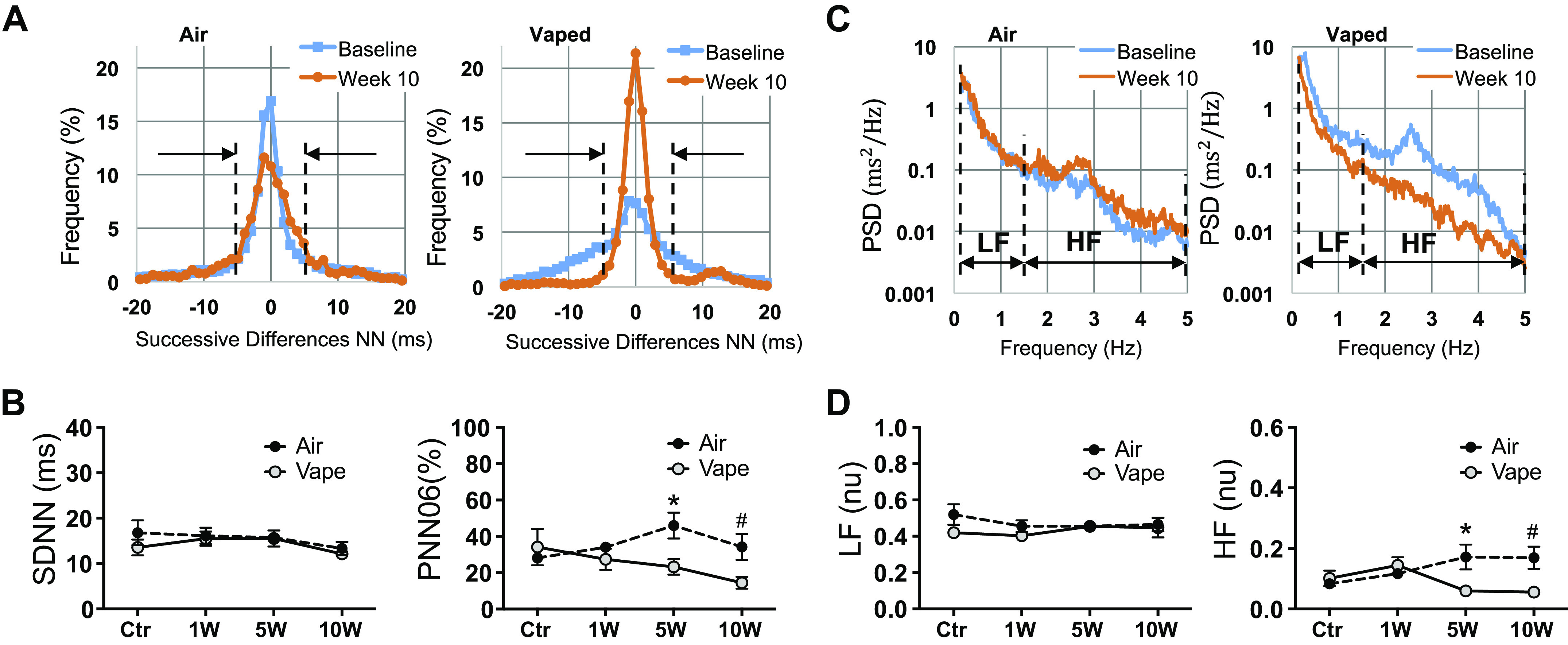
Assessment of heart rate variability in mice exposed to air or vanilla custard e-vapor. A: histograms of the successive differences in NN segments in an air control (left) and a vaped mouse (right) at baseline (blue) and at 10-wk exposure (orange). B: temporal parameters SDNN (ms) and pNN06 (%) at baseline and 1-wk, 5-wk, and 10-wk exposure to air (black circles, n = 5 animals) or vaping (gray circles, n = 5 animals). C: periodograms as estimators of the PSD in an air control (left) and a vaped mouse (right) at baseline (blue) and at 10-wk exposure (orange). D: spectral parameters LF (nu) and HF (nu) at baseline, 1-wk, 5-wk, and 10-wk exposure to air (black circles, n = 5 animals) or vaping (gray circles, n = 5 animals). *P < 0.05, t test, vape vs. air at 5 wk; #P < 0.05, t test, vape vs. air at 10 wk. HF, high frequency; LF, low frequency; SDNN, standard deviation of the normal sinus beats.