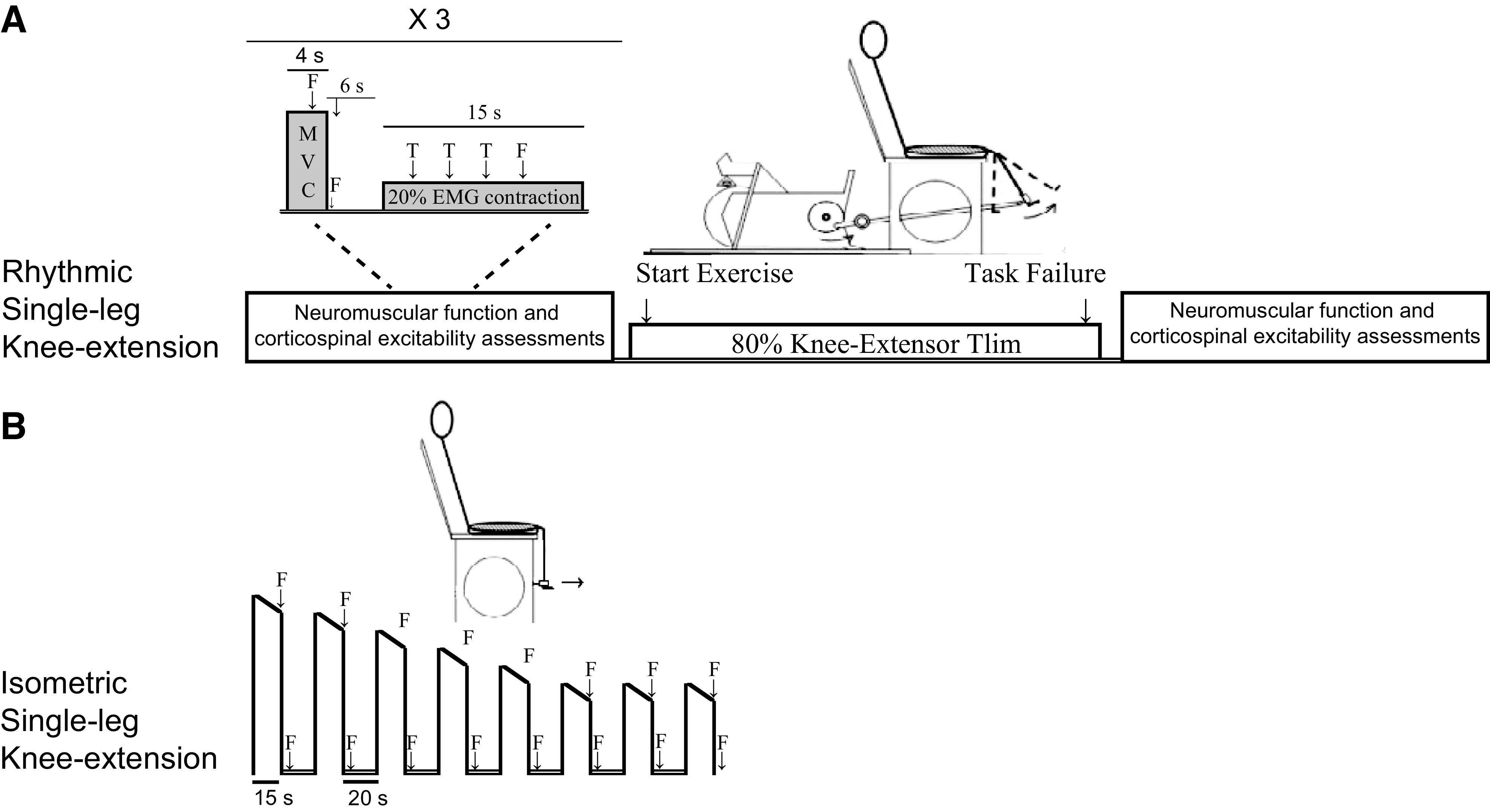
Figure 1.

Schematic illustration of the 2 exercise protocols. Protocol A included the assessment of neuromuscular and corticospinal function before and after exercise. In protocol B, fatigue was quantified during exercise using femoral nerve stimulations (F). EMG, electromyography; MVC, maximal voluntary quadriceps contraction; T, transcranial magnetic stimulation; Tlim, time-to-task failure: 20% EMG contraction, 20% of the EMG attained during the preexercise MVC.
