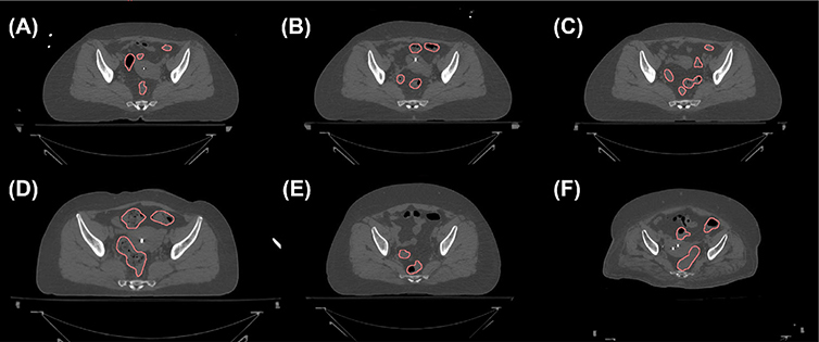
Fig. 1.
Axial CT slices containing the sigmoid colons (contours). Multiple disjointed regions can belong to the same sigmoid colon, as they are connected via the third dimension. (a)–(c) The same patients scanned at three different days separated by about one week. CT slices approximately at the same axial level are shown. (d)–(f) Three other patients.

