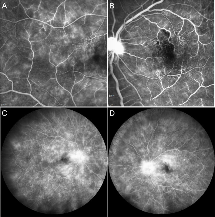
Fig. 2.
Fluorescein Angiography – Bilateral Occlusive Vasculopathy at Presentation. a Fluorescein angiography (FA) of the right eye temporal to the macular showing leakage from retinal capillaries, and teleangiectatic vessels (3.30 min). b FA of the left eye showing hypofluorescence superior and involving the fovea, compatible with hypoperfusion due to branch retinal artery occlusion. Note leakage from retinal capillaries and optic disc (1.18 min). c-d Late phase showing capillary leakage, peripheral capillary nonperfusion and marked hot discs (8 min)

