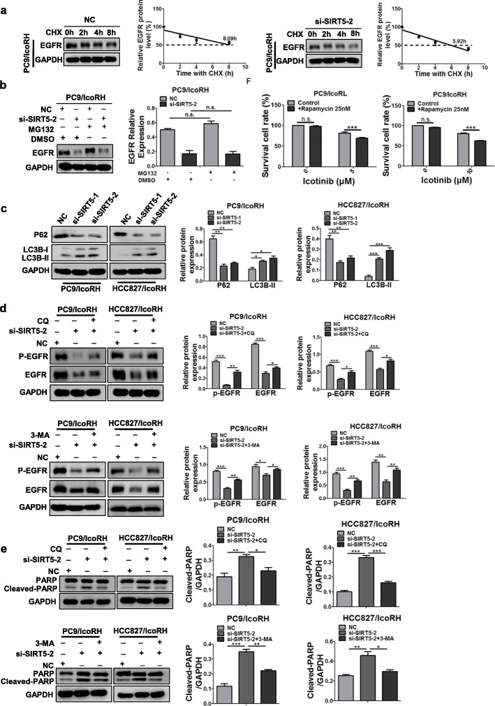
Fig. 6.
Inhibition of autophagic degradation of EGFR by SIRT5. a The protein level of EGFR in PC9/IcoRH cells with the treatment of CHX (20 μg/ml) for 0, 2, 4 and 8 h were determined by western blot. b The protein level of EGFR in SIRT5 KD-PC9/IcoRH cells with the treatment of MG-132 (10 μM) for 12 h were determined by western blot. c The level of LC3B and p62 in SIRT5 KD-icotinib-resistant cells (PC9/IcoRH and HCC827/IcoRH) was determined by western blot. d-e The level of EGFR, p-EGFR (d) and PARP (e) in SIRT5 KD-icotinib-resistant LUAD cells (PC9/IcoRH and HCC827/IcoRH) treated with CQ or 3-MA was determined by western blot. f The icotinib sensitivity in icotinib-resistant LUAD cells (PC9/IcoRL and PC9/IcoRH) with or without rapamycinfor 72 h was determined by MTT assay. The mean ± SD of triplicate experiments were plotted, *P < 0.05, **P < 0.01, ***P < 0.001, n.s., not statistically significant

