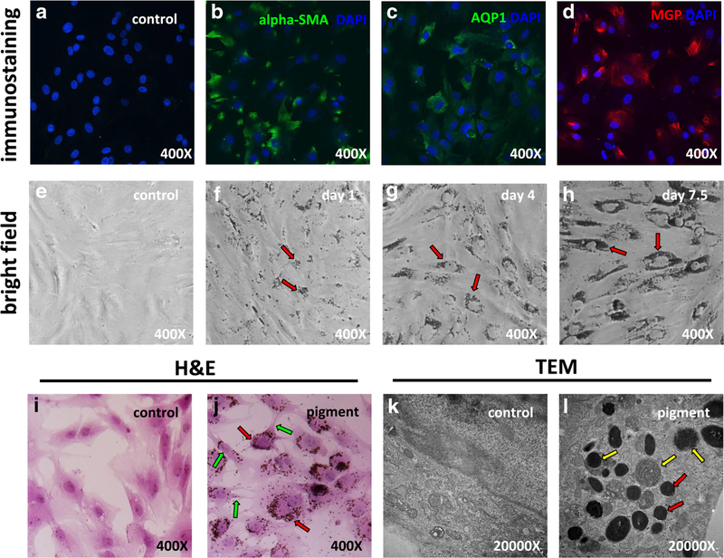
Fig. 1.
Primary trabecular meshwork (TM) cell characterization and pigment exposure. Probing with TM-specific markers alpha-SMA (b), AQP1 (c), and MGP (d) revealed positive staining for all markers. Bright field imaging of TM cells after 1 day (f, red arrows), 4 days (g, red arrows), and 7.5 days (h, red arrows) of pigment showed accumulation of pigment granules in the cytoplasm and around the cell nuclei. H&E staining of TM cells showed that the pigment granules were located within cells (j, red arrows). Some contraction of cell bodies was observed (j, green arrows). Transmission electron microscopy showed the ultrastructure of organelles (k). In the control (c) group, there were no pigment granules within cells, but in the pigment dispersion group, abundant pigment granules were found in the cytoplasm (l, red arrows). Some pigment granules were in different stages of phagolysosomal digestion (l, yellow arrows)

