Fig. 5.
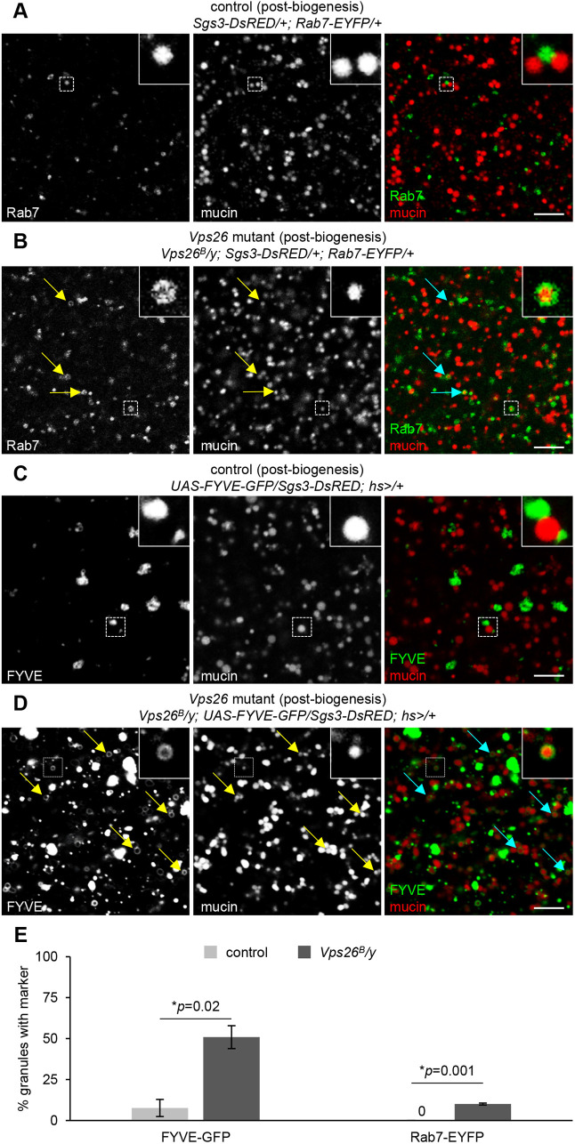
Secretory cargo begins to accumulate within endosomes at biogenesis. (A) Live-cell imaging of Rab7-EYFP (green) and mucins (Sgs3-DsRED; red) in control cells immediately following the onset of secretory granule biogenesis. Rab7 localizes in small puncta that are completely distinct from mucins. Boxed areas are magnified at the upper right of images. (B) Live-cell imaging of Rab7 and mucins in Vps26 mutant cells immediately following the onset of secretory granule biogenesis. Rab7 endosomes appear slightly enlarged compared to controls (A) and some contain mucin cargo, indicated by arrows. Boxed areas are magnified at the upper right of images. (C) Live-cell imaging of the PI3P marker FYVE-GFP (green) and mucins (Sgs3-DsRED; red) in control salivary glands post-biogenesis. FYVE-GFP and mucins are completely distinct; boxed areas are magnified at the upper right of images. (D) Live-cell imaging of FYVE-GFP and mucins in Vps26 mutant salivary glands post-biogenesis. A significant number of mucin-containing vesicles are surrounded by FYVE-GFP, indicated by arrows. Boxed areas are magnified at the upper right of images. Scale bars: 5 µm. (E) Quantification of the percentage of mucin-containing vesicles surrounded by FYVE-GFP or Rab7-EFYP shows a significant increase in Vps26 mutant cells. Note that no mucin-containing vesicles were surrounded by Rab7-EYFP in control cells. Graphs show mean±s.d. of the percentage of mucin-containing vesicles surrounded by FYVE-GFP or Rab7-EYFP in cropped images of three cells from three separate salivary glands for each genotype. Mucin-containing vesicles counted: FYVE-GFP, control n=93; FYVE-GFP, Vps26 mutant n=120; Rab7-EYFP, control n=91; Rab7-EYFP, Vps26 mutant n=79. Statistics calculated by two-tailed paired Student's t-test.

