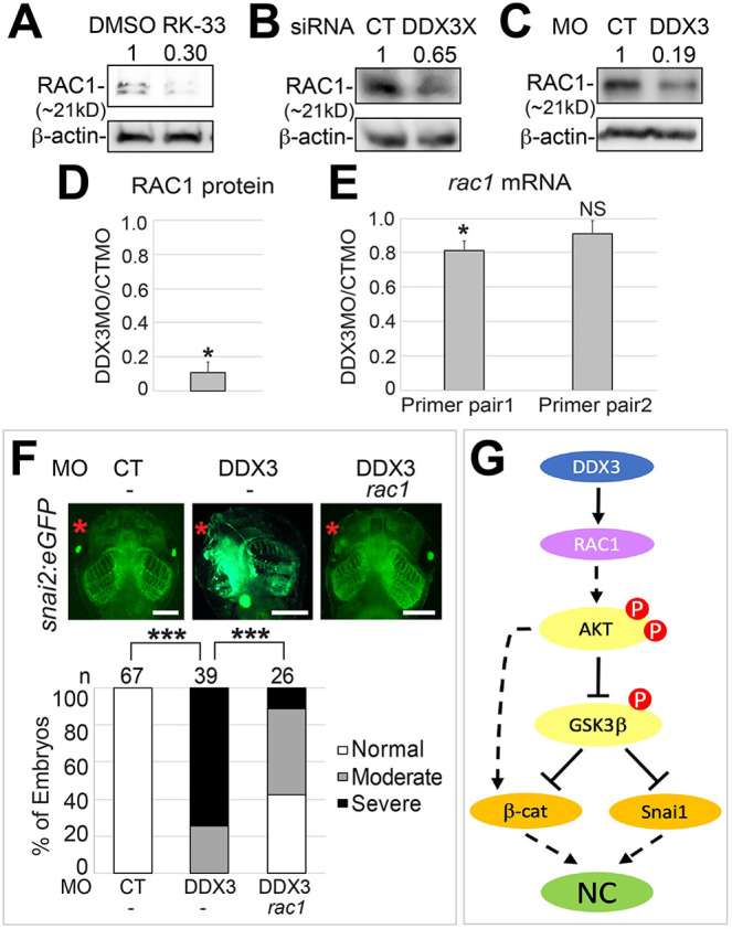
Fig. 6.

RAC1 is a downstream effector of DDX3 in NC induction. (A,B) HEK293T cells were treated with RK-33 (A) or transfected with the indicated siRNA (B) as in Fig. 3. Cell lysates were processed for western blotting for RAC1. (C) Embryo lysates shown in Fig. 3B were reblotted for RAC1. Results of three independent experiments are summarized in D. Density of RAC1 was normalized against that of β-actin, and fold change (DDX3 MO versus control MO) is shown. *P<0.05 (unpaired t-test). (E) The same batches of injected embryos in C and D (three biological replicates) were processed for RT-qPCR using two pairs of primers for rac1 mRNA. Results were normalized against gapdh mRNA (internal control). *P<0.05; NS, not significant (unpaired t-tests). (D,E) Data are mean±s.e.m. (F) One anterodorsal (D1) blastomere of eight-cell stage snai2:eGFP embryos was injected with the indicated MO (1.5 ng each) and mRNA (100 pg). Embryos were cultured to stage ∼46 and imaged for eGFP expression. Representative embryos are shown in ventral view with anterior at the top. n, number of embryos scored. ***P<0.001 (χ2 tests). (G) A model for DDX3 function in regulating downstream signaling during NC induction. Scale bars: 500 μm.
