
Fig. 9. Analysis of the effect of Agno on mitochondrial membrane potential.
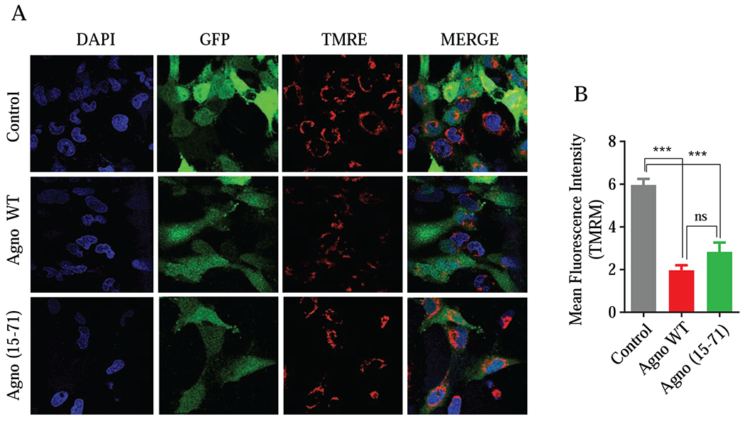
(A) SVGA cells were co-transfected with expression plasmids in the following combination: pGFP-N1 plus pCGT7-Agno WT or pGFP-N1 plus pCGT7-Agno (15-71) mutant in 1:6 ratio using lipofectamine 3000 reagent. At 18 h post-transfection, cells were subjected to flow cytometry to sort GFP-positive cells and subsequently treated with TMRM following the manufacturer’s recommendations as described under materials and methods. The live cells were then imaged by using a Carl Zeiss 510 confocal microscope while maintaining them at 37°C and in a humidified atmosphere with 5 % CO2. (B) Images were quantified using NIH ImageJ software and mean integrated intensities of images (20 randomly chosen fields) in the red channel were determined after background subtraction. Finally, data was presented in a graph form.