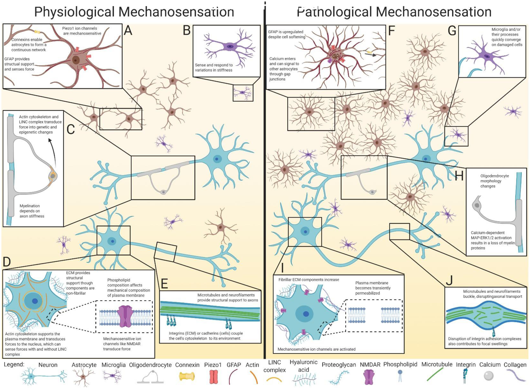
Figure 2:

Physiological and pathological mechanosensation in neurons and glia. A: Astrocytes are linked together in a continuous network of gap junctions formed from connexins. This network may act as a scaffold to sense forces throughout the brain via mechanosensitive Piezo1 channels, along with cytoskeletal elements such as GFAP. B: Microglia can adapt their spread area, morphology, and cytoskeleton to changes in environmental stiffness. C: Oligodendrocytes sense force through their cytoskeleton and LINC complex, resulting in transcriptional changes to promote differentiation and maturation. Once mature, myelination itself depends on the ability to sense the stiffness and diameter of axons. D: Neuronal mechanical properties are affected by the composition of the ECM and plasma membrane. Applied forces are transduced through mechanosensitive components such as ion channels, cytoskeletal proteins, and the nucleus. E: In the axon, cytoskeletal proteins such as microtubules and neurofilaments provide structural support. The cytoskeleton is coupled to the environment via transmembrane proteins such as integrins and cadherins that transduce mechanical forces. F: In response to supra-threshold forces, astrocytes proliferate and become hypertrophic, upregulating components of the cytoskeleton like GFAP, despite becoming softer. The opening of mechanosensitive ion channels such as Piezo1 and NMDAR allow cations including calcium to enter the cell, which may signal to other connected astrocytes through gap junctions. G: Activated microglia and their processes converge on damaged cells and areas of increasing stiffness. H: Oligodendrocytes experiencing high strain display altered morphology and decreased myelin protein expression. Myelin organization can become disrupted, particularly in paranodal regions. I: Neuronal plasma membranes experience small tears, which along with activation of mechanosensitive ion channels, disrupt ion homeostasis. Supra-threshold forces also result in altered ECM composition, with a shift to stiffer substrates. J: Diffuse axonal injury results from the breakage of cytoskeletal components such as microtubules and neurofilaments, which along with disruption of adhesion complexes, results in focal swellings. Although beyond the scope of this graphic, note that mechanosensation pathways are also critical to brain vasculature homeostasis and pathophysiology, as reviewed in Monson et al. (2019), Baeyens et al. (2016), and Fang et al. (2020).
