
Figure 1.
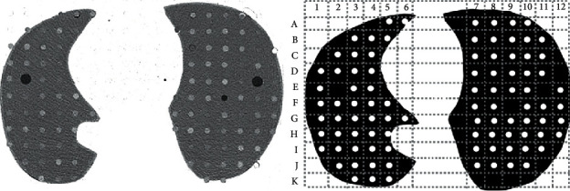
Example of the selected CT slice the radiologists analyzed and the printed diagram in which they registered the nodules they detected. Peripheral nodules such as A3, A4, and B2 were excluded from analysis because of their interface with the chest wall. Positions E2, E11, and F9 had no simulated nodules and served as reference to count columns and rows.