Figure 2.
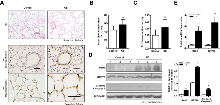
Rcn3 expression in lung tissues from CS-induced mouse model of emphysema. A total of 16 C57BL/6 mice (6 weeks old) were exposed to cigarette smoke for 6 months as emphysema mice (CS group) and 16 C57BL/6 mice were exposed to air for 6 months as Control mice (Control group). (A) Photomicrography of pulmonary parenchyma stained with H&E (upper panels). Bar size: 100 μm; IHC assays of Rcn3 (low and high magnifications, lower panels). Bar size: 50 μm; (B) Morphometric analysis of the MLI ((44.00 ± 2.528 μm vs.56.81 ± 3.053 μm, n = 16 per group, p<0.01); (C) The mean optical density of Rcn3 expression was measured in the lung tissues according to the Rcn3 IHC assays (3 random views, 100× amplification for one subject, n = 16 per group, p<0.01); (D) Representative WB for protein levels of Rcn3, GRP78 and Cleaved caspase-3 in lungs and the ratios to β-Tubulin presented by bar graph, n = 8 per group; (E) Expressions of Rcn3 and GRP78 mRNA were determined with Qualitative PCR analysis, n = 9–10 per group. Data presented as mean ± SEM; *p<0.05, **p<0.01 versus the Control group.

