
Fig. 2.
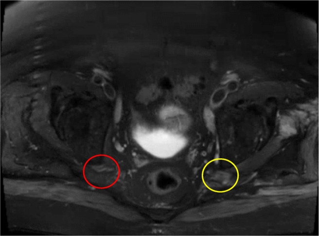
Axial STIR image through the pelvis show asymmetric left focal thickening and increased signal of the sciatic nerve at the level of the posterior acetabular column (yellow circle). The right sciatic nerve appears normal (red circle)
Official websites use .gov
A
.gov website belongs to an official
government organization in the United States.
Secure .gov websites use HTTPS
A lock (
) or https:// means you've safely
connected to the .gov website. Share sensitive
information only on official, secure websites.
Axial STIR image through the pelvis show asymmetric left focal thickening and increased signal of the sciatic nerve at the level of the posterior acetabular column (yellow circle). The right sciatic nerve appears normal (red circle)