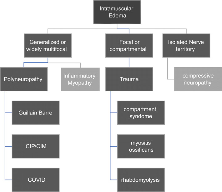
Fig. 3.
A limited flowchart illustrating the approach to differential diagnosis of muscle edema, as seen in this case. Not all possibilities can be included in such a format. For example, radiation-related changes (which could be regarded as a form of trauma) are not included, nor is peritumoral edema

