Fig. 1.
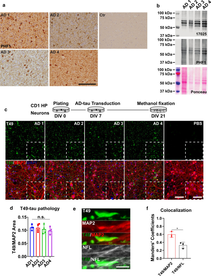
Insoluble tau from different AD cases exhibits consistent seeding abilities in WT mouse primary neurons. a Immunohistochemistry (IHC) of frontal cortex sections from different AD patient brains and non-tau pathology control using the PHF1 anti-phosphorylated-tau antibody; scale bar = 500 µm. b Immunoblot of sarkosyl-insoluble AD-tau probed with a total tau (17,025) and a phospho-tau (PHF1) antibody; ponceau staining shows total protein in the samples. c Immunocytochemistry (ICC) of AD-tau-induced mouse tau pathology in CD1 mouse primary hippocampal neuron cultures revealed by a mouse tau-specific antibody (T49) with concomitant staining of cell nuclei (DAPI) and neuronal dendrites (MAP2 antibody); scale bar = 100 µm and 25 µm (inset). The schematic diagram shows the experimental paradigm for the neuron culture-based activity assay. d Quantification of T49-mouse tau pathology from c. T49-postive area was normalized to the MAP2 positive area; n.s. nonsignificant for all groups; one-way ANOVA followed by Tukey post hoc test; n = 4. e Representative images of T49-positive tau pathology shows the relative location of AD-tau induced mouse tau pathology in dendrites (MAP2) and axons (NFL); scale bar = 5 µm. f Colocalization of T49/MAP2 and T49/NFL from e; n = 3

