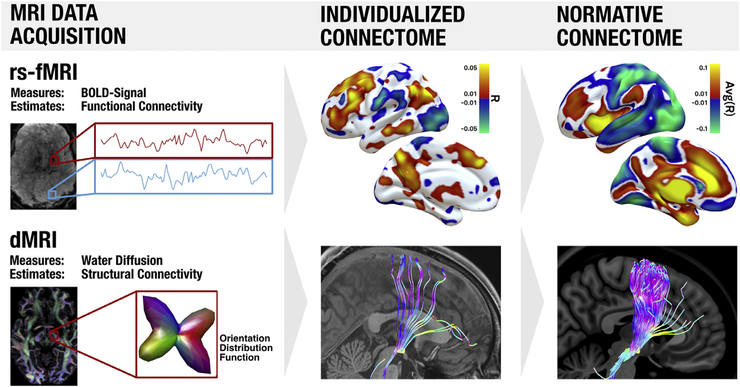
Fig. 2.
Noninvasive MRI based methods to estimate brain connectivity. Top: resting-state functional connectivity MRI (rs-fMRI) is based on spontaneous fluctuations in brain activity as indexed by the blood-oxygen-level-dependent (BOLD) signal. This signal is recorded from all voxels simultaneously, and voxels in which the fluctuations are correlated are considered functionally connected. Areas positively correlated to a seed region (right subthalamic nucleus, red box) are shown in hot colors, while regions negatively correlated (anticorrelated) to the seed region are shown in cool colors. Results based on a single subject are shown in the middle column (individualized connectome) while results based on 1000 subjects are shown in the right column (normative connectome). Bottom: diffusion-weighted imaging (dMRI) measures water diffusion which is anisotropic in the brain. In general, diffusion is stronger along the direction of larger fiber bundles as opposed to orthogonal to them. Based on local diffusion properties of each voxel (which can be represented as orientation distribution functions), tractography algorithms can estimate the location of white-matter bundles to provide an estimate of structural connectivity. White matter bundles passing through the subthalamic nucleusare shown for a single subject in the middle column and for a group of 1000 subjects in the right column. Displayed data are from the human connectome andgenome superstruct projects (Holmes et al., 2015; van Essen and Ugurbil, 2012).

