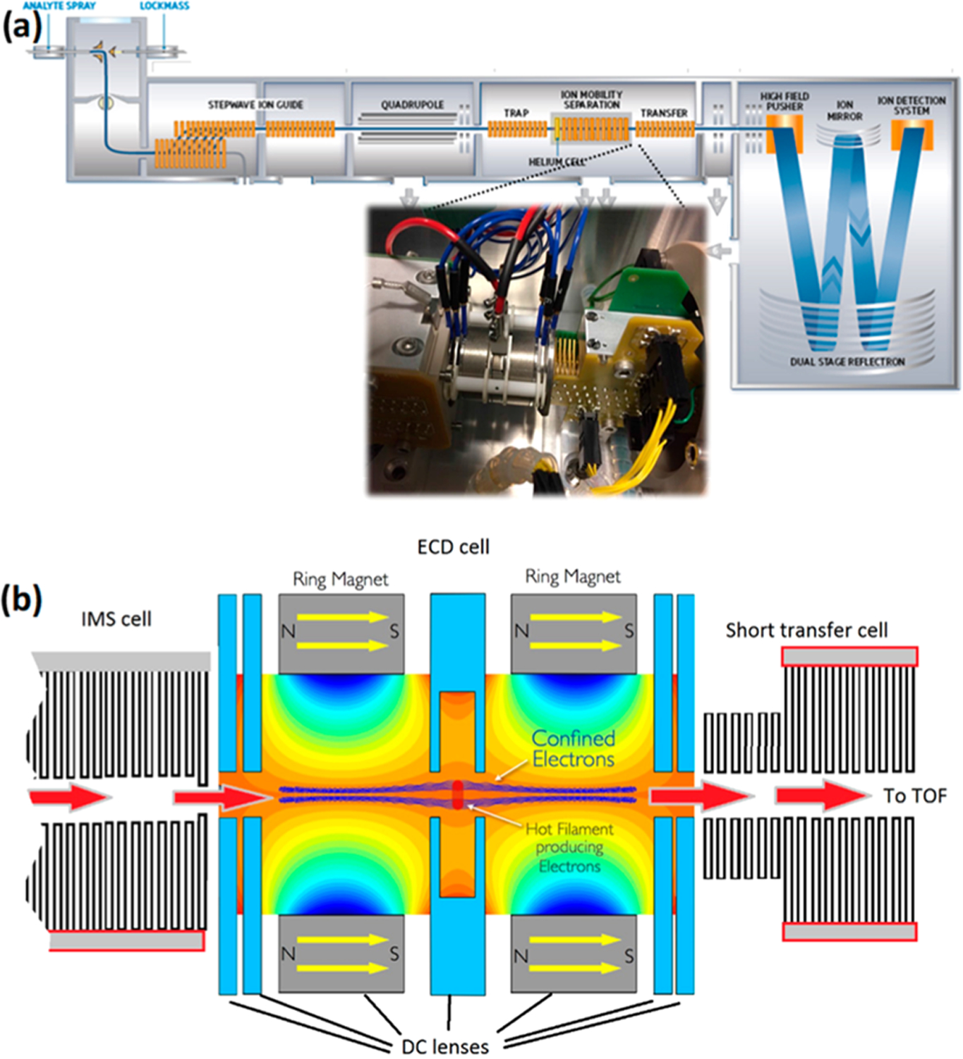
Figure 1.

(a) Schematic representation of the Synapt G2-Si instrument used in this study. The inset shows a photo of the EMS cell mounted between the IM and transfer cell. (b) Schematic of the EMS cell, highlighting the path of the protein ions (red arrows) and confined electrons (in blue).
