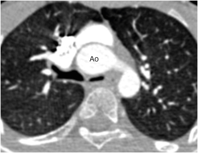
Figure 17. Axial contrast enhanced CT angiography image in a TGA patient after ASO showing compression of the left main bronchus between the ascending aorta and the spine. Note the subtle hypoattenuation of the left lung as compared to the right, likely due to air trapping.
Ao: neo-aorta root, ASO: arterial switch operation, CT: computed tomography, TGA: transposition of the great arteries.

