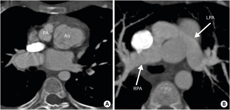
Figure 5. Axial maximum intensity projection CT angiography images showing abnormal great vessel configuration in TGA patient after ASO. (A) Image showing extreme rightward deviation of the neo-pulmonary root with respect to the aorta. (B) Image showing stretching of the LPA around the aorta in the same patient. The LPA is narrower in diameter as compared to right. The excessive rightward relation of the neo-pulmonary root leads to stretching of the LPA, which predisposes the patient to LPA stenosis.
Ao: neo-aorta root, ASO: arterial switch operation, CT: computed tomography, LPA: left pulmonary artery, PA: pulmonary artery, RPA: right pulmonary artery, TGA: transposition of the great arteries.

