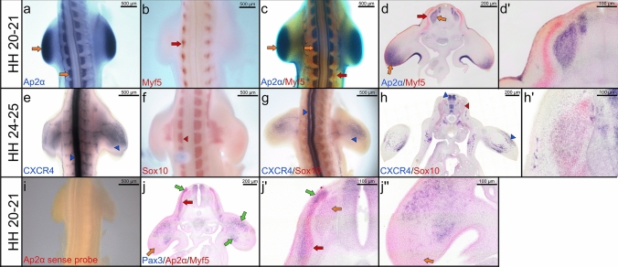
Fig. 1.
Double whole-mount ISH. Representative embryos are shown below: a–c Embryos of a stage HH20-21 are labelled with a DIG- Ap2α probe (blue) and a FITC-Myf5 probe (red). d Vibratome cross sections of double whole-mounted embryos in (c). dʹ Higher magnification of the photo in (d). e–g Embryos of a stage HH24-25 are labelled with a DIG-CXCR4 probe (blue) and a FITC-Sox10 probe (red). h Vibratome cross sections of double whole-mounted embryos in (g). hʹ Higher magnification of the photos in (h). i Chicken embryo of a stage HH22 hybridized with Ap2α sense probe as a negative control. j Vibratome cross sections of double whole-mounted embryos labelled with a FITC- Ap2α/ Myf5 probes (red) and a DIG-Pax3 probe (blue). Scale bar 200 μm. jʹ, jʹʹ Higher magnification of the photos in (j). Orange arrows indicate Ap2α expression; red arrows indicate Myf5 expression; blue arrowheads indicate CXCR4 expression; red arrowheads indicate Sox10 expression; green arrows indicate Pax3 expression. Whole-mount photos were taken at a magnification of 5×. Cross section photos were taken at a magnification of 40×

