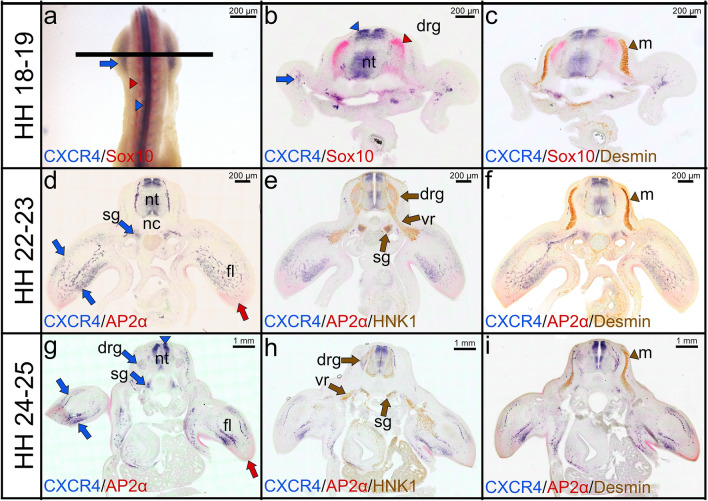
Fig. 3.
Combined double whole-mount ISH and immunostaining labelling for CXCR4, Sox10, Ap2α, HNK1 and Desmin. a Double whole-mounted embryo labelled with CXCR4 probe in blue (DIG), and Sox10 probe in red (FITC). CXCR4 transcripts are detected strongly in the limb bud (blue arrows) and neural tube (blue arrowhead). Sox10 transcripts are expressed in the dorsal root ganglia (red arrowhead). b Vibratome cross sections (indicated by the line in a) showing expression of CXCR4 (blue) and Sox10 (red). c Immunostaining was performed for Desmin on the same cross section in (b). d, g Vibratome cross sections showing expression of CXCR4 (blue) and Ap2α (red). e, h Immunostaining was performed for HNK1 on the same cross section in (d) and (g). f, i Immunostaining was performed for Desmin on the same cross section in (d) and (g). CXCR4 is expressed in the sympathetic ganglia (blue arrows in d, g). The myotome is labelled with Desmin antibody in brown (brown arrowheads in f, i). CXCR4 transcripts and HNK1 antibody are co-expressed in sympathetic ganglia. CXCR4 and Desmin are co-expressed in the limb buds. HNK1 is detected in dorsal root ganglia, ventral roots and sympathetic ganglia (brown arrows in e, h). Ap2α transcripts are expressed in the distal mesenchyme (red arrows) of limb buds. fl forelimb, nt neural tube, nc notochord, drg dorsal root ganglia, vr ventral root, m myotome, sg sympathetic ganglia. Photos are taken with a magnification 40×

