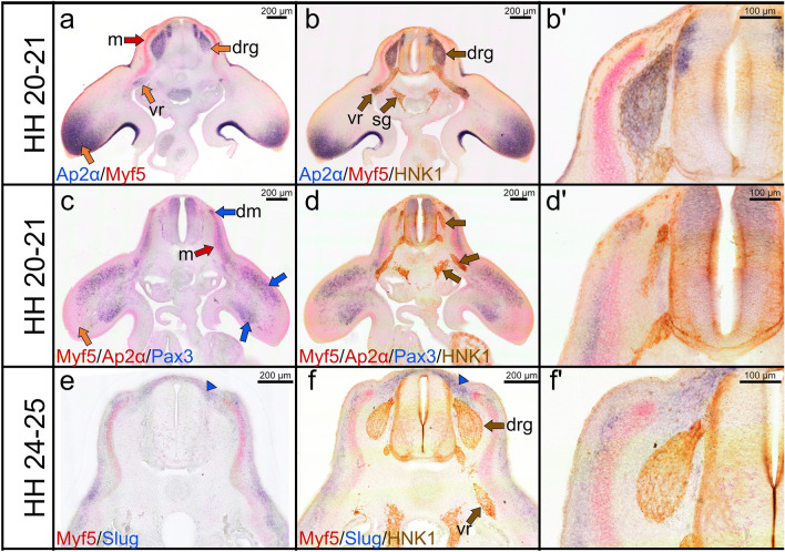
Fig. 5.
Combined double ISH and immunostaining labelling for Myf5, Ap2α, Slug, Pax3 and HNK1. a Cross section of a stage HH20-21 chicken embryo labelled with Ap2α probe in blue and Myf5 probe in red. b Immunostaining is performed for HNK1 on the same cross section as presented in (a). bʹ Higher magnification of the photos in (b). Myf5 signals are found in the myotome (red arrow in a). Ap2α is expressed in the dorsal root ganglia, ventral roots and distal limb bud (orange arrows). The dorsal root ganglia and ventral root are co-labelled with HNK1 in brown colour (brown arrows b). HNK1 is faintly expressed in the neural tube (c) Cross section of a stage HH20-21 stained for Myf5/Ap2α in red (FITC) and Pax3 in blue (DIG). d Immunostaining is performed for HNK1 on the same cross section as presented in (c). dʹ Higher magnification of the photos in (d). Pax3 is expressed in the dermomyotome and migrating muscle progenitor cells in the limb bud (blue arrows c). e Cross section of a stage HH24-25 stained for Myf5 in red (FITC) and Slug in blue (DIG). f Immunostaining is performed for HNK1 on the same cross-section as presented in (e). fʹ Higher magnification of the photo in (f). Note Slug expression in the meninges surrounding the dorsal root ganglia (blue arrowheads). The abbreviations of the cross sections are as indicated before

