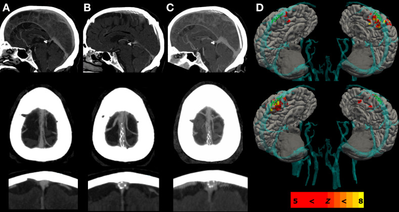
Figure 2.
Pre- and post-neurointervention imaging. Panel A displays the baseline computed tomography venography study of the superior sagittal sinus in sagittal, axial and coronal views for participant 1. Panel B panel displays the repeat study at 3 months, and Panel C at 12 months following implantation of the Stentrode in the superior sagittal sinus, which revealed no evidence of thrombosis, stenosis or device migration. Panel D shows the regions of lower limb blood-oxygen-level-dependent (BOLD) activation relative to cortical and vascular structures derived from a preoperative magnetic resonance imaging study, co-registered to the superior sagittal sinus on intra-operative 3D digital subtraction angiography image.

