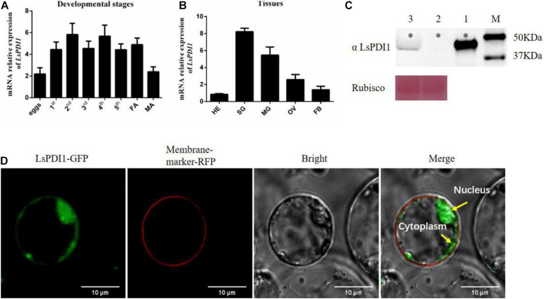
FIGURE 1.
Spatiotemporal expression and subcellular localization of small brown planthopper (SBPH) LsPDI1 in rice protoplast. (A,B) LsPDI1 expression at various developmental stages (A) and in different tissues (B) (n = 5). (C) Western blot detection of LsPDI1 protein (∼48 kDa) secreted in rice plants infested or uninfested by SBPH nymphs (n = 3). Lane 1, salivary glands extracts from SBPH fifth-instar nymphs. Lanes 2 and 3, extracts from rice plants either uninfested (Lane 2) or infested (Lane 3) with fifth-instar nymphs. M, molecular weight marker (kDa); ∗indicates the target band of protein; Rubisco was the loading sample control and Ponceau staining was used. (D) Transient LsPDI1 expression was localized to rice protoplast cytoplasm and nucleus. LsPDI1-GFP was expressed in rice protoplasts by polyethylene glycol-mediated transformation. Fluorescence signals were observed at 488 nm (GFP) and 562 nm (RFP) by confocal laser-scanning microscopy. RFP-marker acts as a membrane location indicator. Bar = 10 μm.

