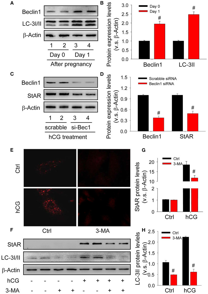
Figure 2.
Autophagy is induced during the luteinization of granulosa cells in vivo and in vitro. (A) The expressions of autophagy marker proteins LC-3I/II and beclin1 on Day 0 or 1 after pregnancy were detected by Western blot. (B) Summarized intensities of StAR blotting normalized to the control (Day 0). (C) Granulosa cells were transfected with siRNA targeting beclin1 for 36 h, and then treated with hCG for 48 h. The expressions of beclin1 and StAR were analyzed by Western blot. (D) Summarized intensities of beclin1 and StAR blotting normalized to the control (scrabble siRNA). (E) Immunofluorescence of LC-3I/II in granulosa cells was pretreated with/without hCG and then treated with/without 3-MA for 24 h. (F) The expression changes of LC-3I/II and beclin1 detected by Western blot in granulosa cells pretreated with/without hCG (15 IU/ml) for 48 h and then treated with 3-MA (10 mM) for 24 h before analysis. (G) Summarized intensities of StAR blotting normalized to the control. (H) Summarized intensities of LC-3II blotting normalized to the control. Each value represents the mean ± SE. One-way analysis of variance (ANOVA) was used to analyze the data, followed by a Tukey's multiple range test. n = 6. Ctrl, vehicle; scrabble, scrabble siRNA; si-Bec1, siRNA targeting beclin1; hCG, human chorionic gonadotropin; 3-MA, 3-methyladenine, #P < 0.05, vs. the group in black.

