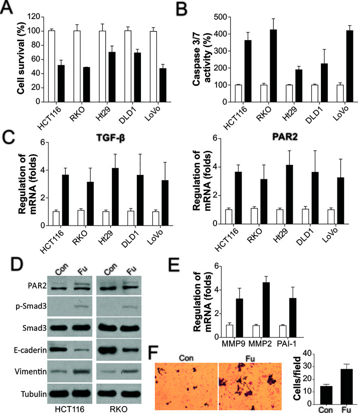
Figure 2.
5-FU treatment induces TGF-β and PAR2 expression and cell migration. (A) The indicated CRC cells were treated with 50 μg/ml of 5-FU for 24 h, and then cell viability was detected using the MTS assay. (B) CRC cells were treated as in (A), and then caspase 3/7 activity was analyzed. (C) CRC cells were processed as in (A), and TGF-β (left) and PAR2 (right) expression was examined using RT-PCR. (D) HCT116 and RKO cells were processed as in (A), after which the expression of the indicated proteins was analyzed through Western blotting. (E) HCT116 cells were processed as in (A), and then the expression of target genes was analyzed using RT-PCR. (F) HCT116 cells were processed as in (A), and the migration of the surviving cells after treatment was analyzed.

