Figure 4.
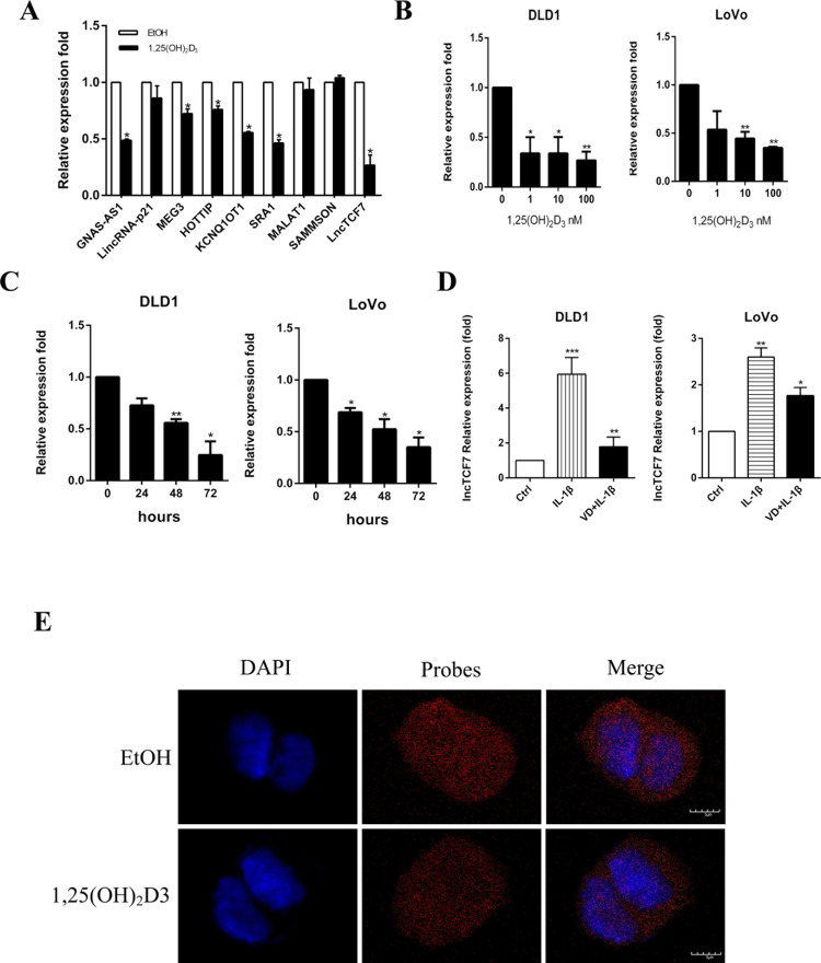
1,25(OH)2D3 inhibits the expression of lncTCF7. (A) Relative mRNA levels of some lncRNAs in DLD1 cells after treatment with 100 nM 1,25(OH)2D3 for 48 h. β-Actin was used as an internal control. (B) The expression level of lncTCF7 in DLD1 and LoVo cells after treatment with increasing doses of 1,25(OH)2D3 for 48 h. β-Actin was used as an internal control. (C) The expression level of lncTCF7 in DLD1 and LoVo cells after treatment with 10 nM 1,25(OH)2D3 for increasing hours. (D) The expression levels of lncTCF7 in DLD1 and LoVo cells were detected by qRT-PCR after treatment with 10 ng/ml IL-1β and 100 nM 1,25(OH)2D3 for 48 h. β-Actin was used as an internal control. (E) LncTCF7 level and localization were studied by RNA-FISH assay on DLD1 cells. After treatment with or without 100 nM 1,25(OH)2D3 for 48 h, DLD1 cells were permeabilized and hybridized with FITC-labeled lncTCF7 probe. Nuclei were stained with DAPI. Red, lncTCF7; blue, nuclei. Scale bar: 5 μm. Data represent the mean ± SEM. *p < 0.05, **p < 0.01, ***p < 0.001.

