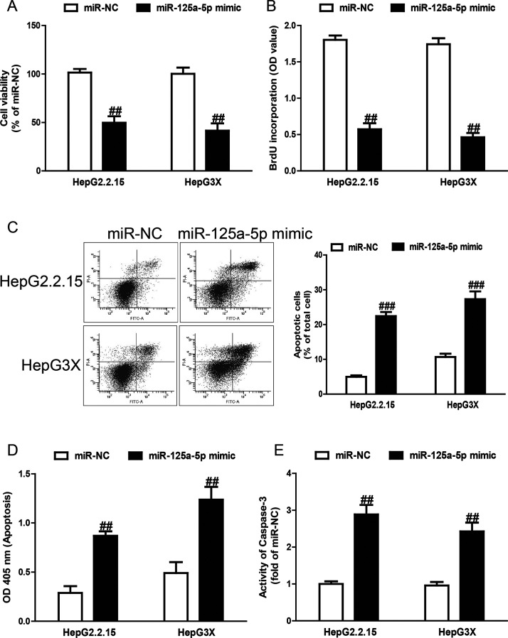
Figure 3.
Effects of miR-125a-5p on cell viability, proliferation, and apoptosis in HepG2.2.15 and HepG3X cells. HepG2.2.15 and HepG3X cells were transfected with miR-125a-5p mimic or miR-NC for 48 h. (A) Cell viability was assessed by cell counting kit-8 (CCK-8) assay. (B) Cell proliferation was assessed by bromodeoxyuridine (BrdU)-enzyme-linked immunosorbent assay (ELISA) assay. Cell apoptosis was measured by flow cytometric analysis of cells labeled with (C) annexin V/propidium iodide (PI) double staining and (D) nucleosomal degradation by using Roche’s Cell Death ELISA Detection Kit, respectively. (E) The activities of caspase 3 were determined by caspase 3 activity detection assay. All data are presented as mean ± SEM, n = 6. ##p < 0.01, ###p < 0.001 versus miR-NC.

