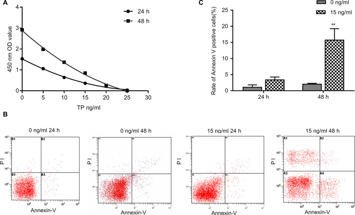
Figure 1.
Effect of triptolide treatment on the cell proliferation and apoptosis of breast cancer cells. (A) MDA-MB-231 cells treated with triptolide (5, 10, 15, 20, and 25 ng/ml) for 24 and 48 h. The level of cell proliferation was detected using the Cell Counting Kit (CCK-8). (B, C) The percentage of apoptotic cells was determined by FACS analysis using annexin V/propidium iodide (PI) staining. Data are presented as the mean ± SD. n = 3. **p < 0.01 versus 0 ng/ml group.

