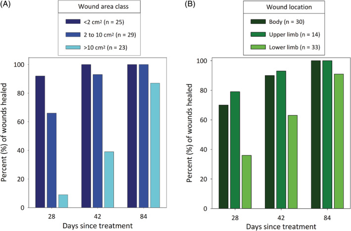
FIGURE 3.

Rate of wound healing as measured by percentages of wounds that had healed by days 28, 42, and 84 after a single treatment with TT (A) in each of 3 maximal wound area classes and (B) at 3 locations (body, upper limb, and lower limb). Data are from the 77 dogs that formed a wound, were assessed at all assessment times through to day 84 after a single treatment with TT, and had complete resolution of the tumor at that time. Data include those dogs with enlarged locoregional LNs detected (metastatic MCT disease could not be confirmed on FNA) at screening. FNA, fine needle aspirate; MCT, mast cell tumor; TT, tigilanol tiglate
