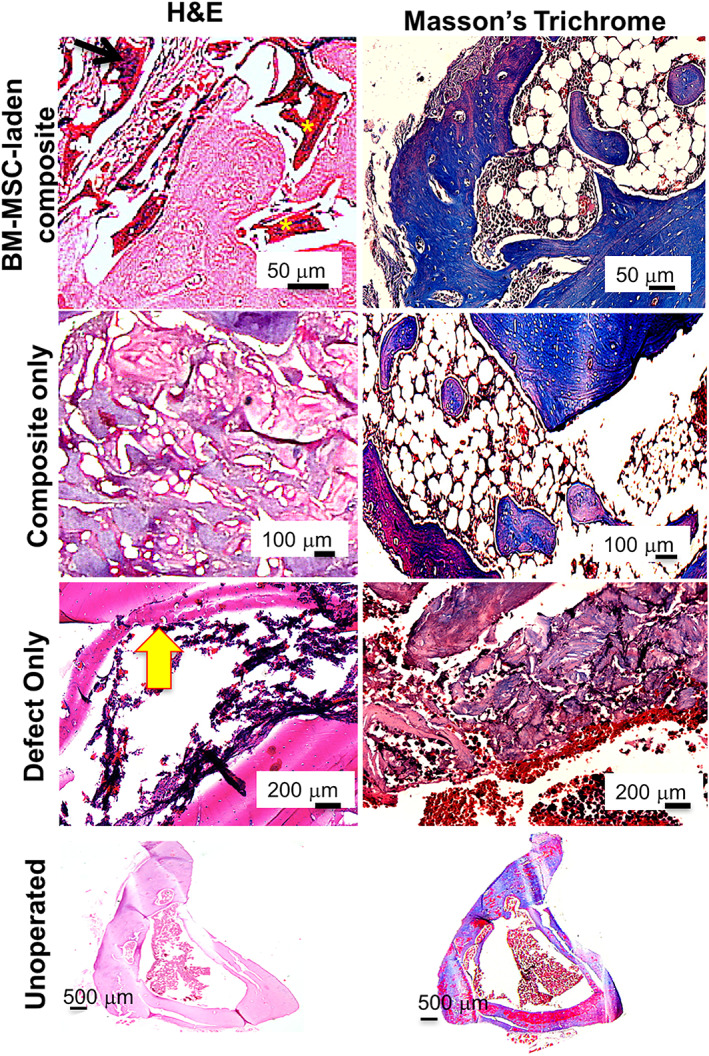
FIGURE 7.

Histology micrographs of ex vivo samples of BM‐MSC‐laden CHT/HA/PCL implanted rat tibiae harvested 6 weeks post surgery. The left side panel depicts H&E staining and Masson's trichrome staining is on the right side panel. CHT, chitosan; HA, hydroxyapatite; H&E, hematoxylin and eosin; PCL, polycaprolactone
