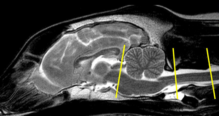
FIGURE 1.

T2‐weighted mid‐sagittal magnetic resonance image of the brain and upper cervical spine of a Beagle. The yellow lines illustrated the location of the phase‐contrast sequence at the mesencephalic aqueduct, the most caudal aspect of the foramen magnum and at the atlantoaxial junction. Measurements were aligned perpendicular to the flow direction
