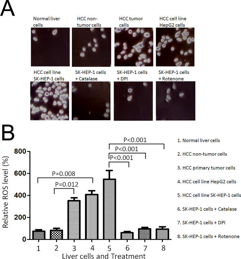
Figure 1.
Hepatocellular cancer cells generated higher levels of endogenous reactive oxygen species (ROS). (A) Normal liver cells THLE-2 cells, hepatocellular carcinoma cells (HCC) nontumor cells, HCC primary tumor cells, and HCC carcinoma cell lines, HepG2 and SK-HEP-1 cells were seeded onto a glass coverslip in a six-well plate at 1 × 10⁵ cells per well for 24 h. CM2-DCFHDA (5 mmol/L) was added into the cell culture medium and incubated for 30 min. For the catalase treatment, catalase (750 U/ml) was added to SK-HEP-1 cells 30 min before the addition of CM2-DCFHDA. The cells were washed three times with 1× PBS and fixed with 10% buffered formalin. The representative images were captured with a confocal fluorescence microscope (at excitation wavelength, 485 nm; emission wavelength, 530 nm). (B) The mean value of DCF fluorescence intensity was obtained from 1 × 10⁵ cells at 485 nm excitation and 540 nm emission settings using a flow cytometer (Becton Dickinson FACSort). SK-HEP-1 cells were treated with solvent, 5 mmol/L DPI, 5 mmol/L rotenone for 30 min, and then stained with 5 mmol/L CM2-DCFHDA for 15 min. The relative fluorescence intensity was analyzed by flow cytometry and normalized to that of normal liver cells. Significant difference was presented when the value of treatment was compared with that of the control.

